
1. Chapter at a Glance
You can easily see the structural similarities and variations in the external morphology of the larger living organisms, both plants and animals. Similarly, if we study the internal structure, we also find several similarities as well as differences. The study of the internal structure of plants is called anatomy. Plants have cells as the basic unit; cells are organised into tissues, and the tissues in turn are organised into organs. Different organs in a plant show differences in their internal structure. Within angiosperms, monocots and dicots are also seen to be anatomically different. This chapter covers plant tissues (meristematic and permanent), the three tissue systems, and the anatomy of dicot and monocot roots, stems and leaves. NEET asks two questions from this chapter almost every year, usually on vascular bundle types, xylem and phloem elements, and dicot vs monocot comparisons.
- Apical, intercalary and lateral meristems; primary vs secondary meristems.
- Parenchyma, collenchyma and sclerenchyma (fibres and sclereids).
- Xylem and phloem elements; protoxylem vs metaxylem; endarch vs exarch.
- Epidermal tissue system: stomata, guard cells (bean-shaped vs dumb-bell shaped), trichomes and root hairs.
- Radial, conjoint closed and conjoint open vascular bundles.
- Comparison of dicot and monocot root, stem and leaf; bulliform cells; Casparian strips.
2. The Tissues
A tissue is a group of cells having a common origin and usually performing a common function. A plant is made up of different kinds of tissues. Tissues are classified into two main groups, namely meristematic and permanent tissues, based on whether the cells being formed are capable of dividing or not.
Meristematic tissues
Growth in plants is largely restricted to specialised regions of active cell division called meristems (Gk. meristos: divided).
- Apical meristems occur at the tips of roots and shoots and produce primary tissues. The root apical meristem occupies the tip of a root, while the shoot apical meristem occupies the distant-most region of the stem axis. During the formation of leaves and elongation of stem, some cells "left behind" from the shoot apical meristem constitute the axillary bud. Such buds are present in the axils of leaves and are capable of forming a branch or a flower.
- Intercalary meristems occur between mature tissues. They are present in grasses and regenerate parts removed by grazing herbivores.
- Since both apical and intercalary meristems appear early in life and contribute to the formation of the primary plant body, they are primary meristems.
- The meristem that occurs in the mature regions of roots and shoots of many plants, particularly those that produce woody axis, and appears later than the primary meristem, is called the secondary or lateral meristem. They are cylindrical meristems. Fascicular vascular cambium, interfascicular cambium and cork cambium are examples of lateral meristems. These are responsible for producing the secondary tissues.
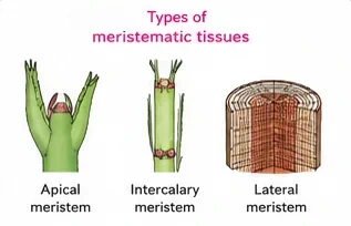
Following divisions of cells in both primary and secondary meristems, the newly formed cells become structurally and functionally specialised and lose the ability to divide. Such cells are termed permanent or mature cells and constitute the permanent tissues.
Permanent tissues
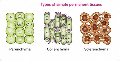
The cells of the permanent tissues do not generally divide further. Permanent tissues having all cells similar in structure and function are called simple tissues. Permanent tissues having many different types of cells are called complex tissues.
| Simple tissue | Cell features | Function | NEET Note |
|---|---|---|---|
| Parenchyma | Forms the major component within organs. Cells isodiametric — spherical, oval, round, polygonal or elongated; walls thin, made of cellulose; cells closely packed or with small intercellular spaces | Photosynthesis, storage, secretion | Living |
| Collenchyma | Occurs in layers below the epidermis in dicotyledonous plants; found either as a homogeneous layer or in patches; cells much thickened at the corners due to a deposition of cellulose, hemicellulose and pectin; oval, spherical or polygonal; often contain chloroplasts; no intercellular spaces | Mechanical support to growing parts such as young stem and petiole of a leaf; assimilate food when they contain chloroplasts | Living; absent in monocots |
| Sclerenchyma | Long, narrow cells with thick and lignified cell walls having a few or numerous pits; usually dead and without protoplasts | Mechanical support to organs | Two types: fibres and sclereids |
- Fibres are thick-walled, elongated and pointed cells, generally occurring in groups in various parts of the plant.
- Sclereids are spherical, oval or cylindrical, highly thickened dead cells with very narrow cavities (lumen). These are commonly found in the fruit walls of nuts; pulp of fruits like guava, pear and sapota; seed coats of legumes and leaves of tea.
Complex tissues: xylem and phloem
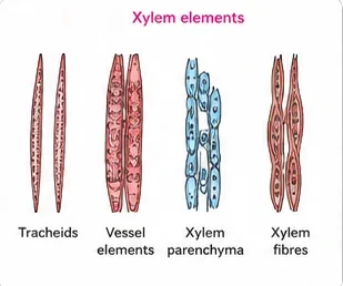
Xylem functions as a conducting tissue for water and minerals from roots to the stem and leaves. It also provides mechanical strength to the plant parts. It is composed of four different kinds of elements, namely tracheids, vessels, xylem fibres and xylem parenchyma.
- Tracheids are elongated or tube-like cells with thick and lignified walls and tapering ends. These are dead and without protoplasm. The inner layers of the cell walls have thickenings which vary in form.
- In flowering plants, tracheids and vessels are the main water-transporting elements. A vessel is a long cylindrical tube-like structure made up of many cells called vessel members, each with lignified walls and a large central cavity. The vessel cells are also devoid of protoplasm. Vessel members are interconnected through perforations in their common walls. The presence of vessels is a characteristic feature of angiosperms.
- Xylem fibres have highly thickened walls and obliterated central lumens. These may either be septate or aseptate.
- Xylem parenchyma cells are living and thin-walled, and their cell walls are made up of cellulose. They store food materials in the form of starch or fat, and other substances like tannins. The radial conduction of water takes place by the ray parenchymatous cells.
- Primary xylem is of two types — protoxylem and metaxylem. The first-formed primary xylem elements are called protoxylem and the later-formed primary xylem is called metaxylem. In stems, the protoxylem lies towards the centre (pith) and the metaxylem lies towards the periphery of the organ. This type of primary xylem is called endarch. In roots, the protoxylem lies towards the periphery and metaxylem lies towards the centre. Such arrangement of primary xylem is called exarch.
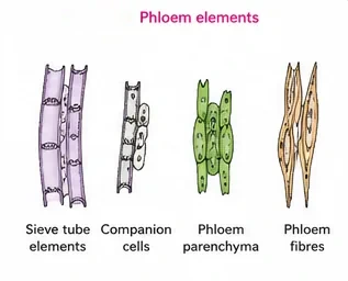
Phloem transports food materials, usually from leaves to other parts of the plant. Phloem in angiosperms is composed of sieve tube elements, companion cells, phloem parenchyma and phloem fibres. Gymnosperms have albuminous cells and sieve cells; they lack sieve tubes and companion cells.
- Sieve tube elements are long, tube-like structures, arranged longitudinally and associated with the companion cells. Their end walls are perforated in a sieve-like manner to form the sieve plates. A mature sieve element possesses a peripheral cytoplasm and a large vacuole but lacks a nucleus. The functions of sieve tubes are controlled by the nucleus of companion cells.
- The companion cells are specialised parenchymatous cells, which are closely associated with sieve tube elements. The sieve tube elements and companion cells are connected by pit fields present between their common longitudinal walls. The companion cells help in maintaining the pressure gradient in the sieve tubes.
- Phloem parenchyma is made up of elongated, tapering cylindrical cells which have dense cytoplasm and nucleus. The cell wall is composed of cellulose and has pits through which plasmodesmatal connections exist between the cells. The phloem parenchyma stores food material and other substances like resins, latex and mucilage. Phloem parenchyma is absent in most monocotyledons.
- Phloem fibres (bast fibres) are made up of sclerenchymatous cells. These are generally absent in the primary phloem but are found in the secondary phloem. These are much elongated, unbranched and have pointed, needle-like apices. The cell wall of phloem fibres is quite thick. At maturity, these fibres lose their protoplasm and become dead. Phloem fibres of jute, flax and hemp are used commercially.
- The first-formed primary phloem consists of narrow sieve tubes and is referred to as protophloem, and the later-formed phloem has bigger sieve tubes and is referred to as metaphloem.
| Element | Living / dead | Main role | NEET Note |
|---|---|---|---|
| Tracheid, vessel | Dead | Water conduction | Vessels typical of angiosperms; absent in most gymnosperms |
| Xylem fibre | Dead | Support | — |
| Xylem parenchyma | Living | Storage; radial conduction | Only living xylem element |
| Sieve tube element | Living, no nucleus | Food conduction | Controlled by companion cell nucleus |
| Companion cell | Living | Maintains pressure gradient | Absent in gymnosperms (albuminous cells instead) |
| Phloem parenchyma | Living | Storage | Absent in most monocots |
| Phloem fibre | Dead | Support | Only dead phloem element; jute, flax, hemp |
In xylem, only xylem parenchyma is living. In phloem, only phloem fibres are dead. A sieve tube element is living but has no nucleus. These three statements produce a question in most years in some form.
3. The Tissue Systems
On the basis of their structure and location, there are three types of tissue systems: the epidermal tissue system, the ground or fundamental tissue system and the vascular or conducting tissue system.
Epidermal tissue system
- The epidermal tissue system forms the outer-most covering of the whole plant body and comprises epidermal cells, stomata and the epidermal appendages — the trichomes and hairs.
- The epidermis is the outermost layer of the primary plant body. It is made up of elongated, compactly arranged cells, which form a continuous layer. Epidermis is usually single-layered. Epidermal cells are parenchymatous with a small amount of cytoplasm lining the cell wall and a large vacuole.
- The outside of the epidermis is often covered with a waxy thick layer called the cuticle, which prevents the loss of water. Cuticle is absent in roots.
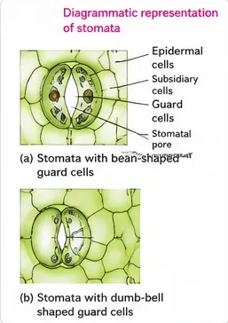
- Stomata are structures present in the epidermis of leaves. Stomata regulate the process of transpiration and gaseous exchange. Each stoma is composed of two bean-shaped cells known as guard cells, which enclose the stomatal pore. In grasses, the guard cells are dumb-bell shaped. The outer walls of guard cells (away from the stomatal pore) are thin, and the inner walls (towards the stomatal pore) are highly thickened. The guard cells possess chloroplasts and regulate the opening and closing of stomata.
- Sometimes, a few epidermal cells in the vicinity of the guard cells become specialised in their shape and size and are known as subsidiary cells. The stomatal aperture, guard cells and the surrounding subsidiary cells are together called the stomatal apparatus.
- The cells of the epidermis bear a number of hairs. The root hairs are unicellular elongations of the epidermal cells and help absorb water and minerals from the soil. On the stem the epidermal hairs are called trichomes. The trichomes in the shoot system are usually multicellular. They may be branched or unbranched and soft or stiff. They may even be secretory. The trichomes help in preventing water loss due to transpiration.
Ground tissue system
All tissues except epidermis and vascular bundles constitute the ground tissue. It consists of simple tissues such as parenchyma, collenchyma and sclerenchyma. Parenchymatous cells are usually present in cortex, pericycle, pith and medullary rays in the primary stems and roots. In leaves, the ground tissue consists of thin-walled chloroplast-containing cells and is called mesophyll.
Vascular tissue system
The vascular system consists of complex tissues, the phloem and the xylem. The xylem and phloem together constitute vascular bundles.
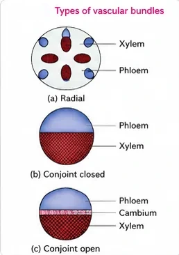
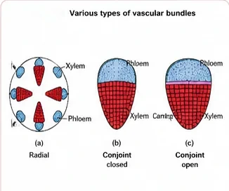
- In dicotyledonous stems, cambium is present between phloem and xylem. Such vascular bundles, because of the presence of cambium, possess the ability to form secondary xylem and phloem tissues, and hence are called open vascular bundles.
- In monocotyledons, the vascular bundles have no cambium in them. Hence, since they do not form secondary tissues, they are referred to as closed.
- When xylem and phloem within a vascular bundle are arranged in an alternate manner along the different radii, the arrangement is called radial, as in roots.
- In conjoint type of vascular bundles, the xylem and phloem are jointly situated along the same radius of vascular bundles. Such vascular bundles are common in stems and leaves. The conjoint vascular bundles usually have the phloem located only on the outer side of xylem.
| Bundle type | Arrangement | Found in | NEET Note |
|---|---|---|---|
| Radial | Xylem and phloem on different radii, alternating | Roots | Exarch xylem |
| Conjoint open | Xylem and phloem on same radius, cambium between them | Dicot stems | Can form secondary tissues |
| Conjoint closed | Xylem and phloem on same radius, no cambium | Monocot stems, leaves | No secondary growth |
4. Anatomy of Roots
Dicotyledonous root (e.g. sunflower)
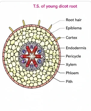
- The outermost layer is the epiblema. Many of the cells of epiblema protrude in the form of unicellular root hairs.
- The cortex consists of several layers of thin-walled parenchyma cells with intercellular spaces.
- The innermost layer of the cortex is called endodermis. It comprises a single layer of barrel-shaped cells without any intercellular spaces. The tangential as well as radial walls of the endodermal cells have a deposition of water-impermeable, waxy material, suberin, in the form of casparian strips.
- Next to endodermis lie a few layers of thick-walled parenchymatous cells referred to as pericycle. Initiation of lateral roots and vascular cambium during the secondary growth takes place in these cells.
- The pith is small or inconspicuous. The parenchymatous cells which lie between the xylem and the phloem are called conjunctive tissue.
- There are usually two to four xylem and phloem patches (diarch to tetrarch). Later, a cambium ring develops between the xylem and phloem.
- All tissues on the inner side of the endodermis, such as pericycle, vascular bundles and pith, constitute the stele.
Monocotyledonous root (e.g. maize)
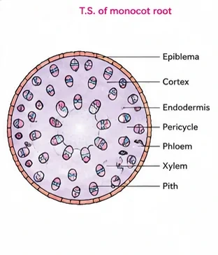
The anatomy of the monocot root is similar to the dicot root in many respects. It has epidermis, cortex, endodermis, pericycle, vascular bundles and pith. As compared to the dicot root, which has fewer xylem bundles, there are usually more than six (polyarch) xylem bundles in the monocot root. The pith is large and well developed. Monocotyledonous roots do not undergo any secondary growth.
5. Anatomy of Stems
Dicotyledonous stem (e.g. sunflower)
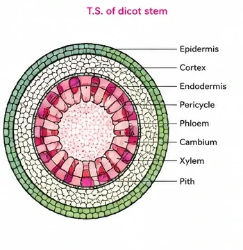
- The transverse section of a typical young dicotyledonous stem shows that the epidermis is the outermost protective layer of the stem. Covered with a thin layer of cuticle, it may bear trichomes and a few stomata.
- The cells arranged in multiple layers between epidermis and pericycle constitute the cortex. It consists of three sub-zones. The outer hypodermis consists of a few layers of collenchymatous cells just below the epidermis, which provide mechanical strength to the young stem. Cortical layers below hypodermis consist of rounded thin-walled parenchymatous cells with conspicuous intercellular spaces. The innermost layer of the cortex is called the endodermis. The cells of the endodermis are rich in starch grains, and the layer is also referred to as the starch sheath.
- Pericycle is present on the inner side of the endodermis and above the phloem in the form of semi-lunar patches of sclerenchyma.
- In between the vascular bundles there are a few layers of radially placed parenchymatous cells, which constitute medullary rays.
- A large number of vascular bundles are arranged in a ring; the "ring" arrangement of vascular bundles is a characteristic of dicot stem. Each vascular bundle is conjoint, open and with endarch protoxylem.
- A large number of rounded, parenchymatous cells with large intercellular spaces which occupy the central portion of the stem constitute the pith.
Monocotyledonous stem (e.g. maize)
- The monocot stem has a sclerenchymatous hypodermis, a large number of scattered vascular bundles, each surrounded by a sclerenchymatous bundle sheath, and a large, conspicuous parenchymatous ground tissue.
- Vascular bundles are conjoint and closed. Peripheral vascular bundles are generally smaller than the centrally located ones.
- The phloem parenchyma is absent, and water-containing cavities are present within the vascular bundles.
| Feature | Dicot root | Monocot root | Dicot stem | Monocot stem |
|---|---|---|---|---|
| Hypodermis | Absent | Absent | Collenchymatous | Sclerenchymatous |
| Vascular bundles | Radial, 2–4 (di- to tetrarch) | Radial, more than 6 (polyarch) | Conjoint, open, in a ring | Conjoint, closed, scattered, with bundle sheath |
| Xylem | Exarch | Exarch | Endarch | Endarch |
| Pith | Small or absent | Large | Large | Not distinct (ground tissue) |
| Secondary growth | Occurs | Absent | Occurs | Absent |
6. Anatomy of Leaves
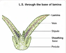
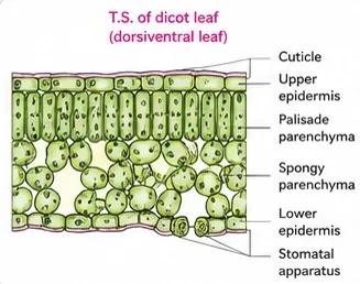
Dorsiventral (dicotyledonous) leaf
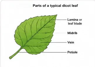
The vertical section of a dorsiventral leaf through the lamina shows three main parts, namely epidermis, mesophyll and vascular system.
- The epidermis, which covers both the upper surface (adaxial epidermis) and lower surface (abaxial epidermis) of the leaf, has a conspicuous cuticle. The abaxial epidermis generally bears more stomata than the adaxial epidermis. The latter may even lack stomata.
- The tissue between the upper and the lower epidermis is called the mesophyll. Mesophyll, which possesses chloroplasts and carries out photosynthesis, is made up of parenchyma. It has two types of cells — the palisade parenchyma and the spongy parenchyma. The adaxially placed palisade parenchyma is made up of elongated cells, which are arranged vertically and parallel to each other. The oval or round and loosely arranged spongy parenchyma is situated below the palisade cells and extends to the lower epidermis. There are numerous large spaces and air cavities between these cells.
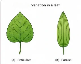
- The vascular system includes vascular bundles, which can be seen in the veins and the midrib. The size of the vascular bundles is dependent on the size of the veins. The veins vary in thickness in the reticulate venation of the dicot leaves. The vascular bundles are surrounded by a layer of thick-walled bundle sheath cells.
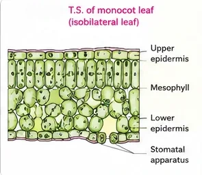
Isobilateral (monocotyledonous) leaf
The anatomy of the isobilateral leaf is similar to that of the dorsiventral leaf in many ways. It shows the following characteristic differences. In an isobilateral leaf, the stomata are present on both surfaces of the epidermis, and the mesophyll is not differentiated into palisade and spongy parenchyma. In grasses, certain adaxial epidermal cells along the veins modify themselves into large, empty, colourless cells. These are called bulliform cells. When the bulliform cells in the leaves have absorbed water and are turgid, the leaf surface is exposed. When they are flaccid due to water stress, they make the leaves curl inwards to minimise water loss. The parallel venation in monocot leaves is reflected in the near similar sizes of vascular bundles (except in main veins) as seen in vertical sections of the leaves.
| Feature | Dorsiventral (dicot) leaf | Isobilateral (monocot) leaf | NEET Note |
|---|---|---|---|
| Stomata | More on lower surface | Nearly equal on both surfaces | — |
| Mesophyll | Palisade + spongy | Undifferentiated | — |
| Bulliform cells | Absent | Present (grasses) | Cause leaf rolling in water stress |
| Vascular bundles | Vary in size (reticulate) | Similar size (parallel) | — |
Bulliform cells are large, empty, colourless adaxial epidermal cells of grass leaves; when they lose water the leaf rolls inward to reduce transpiration. Casparian strips are suberin bands on the radial and tangential walls of root endodermis; they block the apoplast path and force water to pass through cell membranes. Both are favourite statement-type questions.
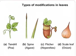
Modifications of leaves (for revision with Morphology)
7. Extra Reading: Secondary Growth
The section on secondary growth was removed in the rationalised NCERT textbook and is not in the current NMC syllabus. It is given briefly here because the pictures are useful and the topic links to "open vascular bundles" and "lateral meristems", which are still in the syllabus.
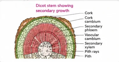
- In dicot stems, the cambium within the bundles (intrafascicular) and the cells of medullary rays next to it (interfascicular cambium) join to form a continuous cambial ring. It cuts off secondary xylem towards the inside and secondary phloem towards the outside. More xylem than phloem is formed.
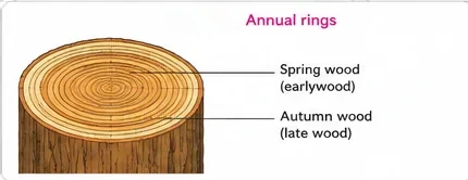
- Cambial activity varies with season. In spring it forms many wide vessels (spring wood or early wood, light in colour, low density); in winter it forms fewer, narrow vessels (autumn wood or late wood, dark, high density). One spring ring plus one autumn ring forms an annual ring; counting rings estimates the age of a tree (dendrochronology).
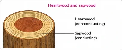
- In old trees, the central secondary xylem becomes dark because of tannins, resins, oils, gums and aromatic substances; this heartwood is non-conducting but durable and resistant to microbes and insects. The peripheral, lighter-coloured sapwood conducts water and minerals.
- The cork cambium (phellogen) develops in the cortex and cuts off cork (phellem) outside and secondary cortex (phelloderm) inside. Phellogen + phellem + phelloderm = periderm. All tissues outside the vascular cambium form the bark. Lenticels allow gaseous exchange through the cork.
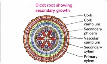
- In dicot roots, the vascular cambium is completely secondary in origin: it starts from conjunctive tissue below the phloem and from pericycle opposite the protoxylem, forming a wavy ring that later becomes circular.
8. Bridge to Class 12: The Ovule
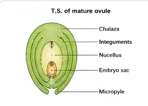
The internal structure of an ovule is studied in detail in Class 12 (Sexual Reproduction in Flowering Plants). The ovule consists of a mass of tissue, the nucellus, enclosed by one or two protective integuments, which leave a small opening called the micropyle. Opposite the micropyle is the chalaza, the basal part. The embryo sac (female gametophyte) lies inside the nucellus.
9. Master Summary Table
| Topic | Key point | NEET Note |
|---|---|---|
| Meristems | Apical, intercalary (primary); lateral (secondary) | Intercalary in grasses |
| Collenchyma | Corner thickenings; living | Absent in monocots |
| Sclerenchyma | Fibres and sclereids; dead, lignified | Sclereids in guava pulp, nut shells |
| Xylem | Tracheids, vessels, fibres, parenchyma | Endarch (stem), exarch (root) |
| Phloem | Sieve tubes, companion cells, parenchyma, fibres | Sieve tube lacks nucleus |
| Stomata | Bean-shaped or dumb-bell (grasses) guard cells | Inner walls thick |
| Vascular bundles | Radial, conjoint open, conjoint closed | Open = cambium present |
| Endodermis | Casparian strips (suberin) | Starch sheath in stem |
| Leaves | Dorsiventral vs isobilateral | Bulliform cells in grasses |
10. Mind Map
- Meristematic
- Simple permanent
- Xylem, phloem
- Epidermal (stomata, hairs)
- Ground
- Vascular bundles
- Dicot root, monocot root
- Dicot stem
- Monocot stem
- Dorsiventral
- Isobilateral
- Bulliform cells
11. Tips and Tricks
- "Root Exarch, Stem Endarch" → R-E, S-E (Exarch = protoxylem outside, towards the Edge).
- Hypodermis: dicot stem = Collenchyma (Curved dicot, soft), monocot stem = Sclerenchyma (Stiff grass).
- Number of xylem bundles: dicot root 2–4 ("few"), monocot root > 6 ("many").
- Monocot stem in one line: "scattered, sheathed, closed, cavities, no phloem parenchyma".
- Saying the sieve tube element has a nucleus (it does not at maturity).
- Saying monocot stems have open bundles (they are closed).
- Saying the outer walls of guard cells are thick (the inner walls, towards the pore, are thick).
- Thinking cuticle is present on roots (it is absent).
- Thinking pericycle in dicot stem is parenchymatous like in roots (in stems it forms semi-lunar sclerenchyma patches).
12. Important One-liners
- Intercalary meristems help grasses regrow after grazing.
- Fascicular cambium, interfascicular cambium and cork cambium are lateral meristems.
- Collenchyma has cellulose, hemicellulose and pectin thickenings at the corners.
- Vessels are characteristic of angiosperms.
- Gymnosperms have sieve cells and albuminous cells instead of sieve tubes and companion cells.
- Phloem fibres of jute, flax and hemp are used commercially.
- Guard cells of grasses are dumb-bell shaped.
- Endodermis of the stem is called the starch sheath.
- Lateral roots arise from the pericycle.
- Monocot stem vascular bundles have water-containing cavities.
- Mesophyll is the ground tissue of a leaf.
- Bulliform cells help grass leaves roll during water stress.
13. NEET-pattern Questions with Detailed Answers
These questions are original and based on concepts that NEET UG asks again and again.
The only living element of xylem is:
- Tracheid
- Vessel
- Xylem parenchyma
- Xylem fibre
Show solution
Xylem parenchyma cells are living and store food. Tracheids, vessels and xylem fibres are dead at maturity.
Answer: (C)
Intercalary meristems are found in:
- Root tips
- Grasses, between mature tissues
- Cork cambium
- Dicot leaves only
Show solution
Intercalary meristems lie between mature tissues, typically at the bases of internodes in grasses. Root tips have apical meristems, and cork cambium is a lateral meristem.
Answer: (B)
Vascular bundles that are conjoint, closed and scattered with a sclerenchymatous bundle sheath are found in:
- Dicot root
- Dicot stem
- Monocot stem
- Monocot root
Show solution
The monocot stem has scattered, conjoint, closed bundles, each with a sclerenchymatous sheath. Roots have radial bundles, and dicot stems have open bundles arranged in a ring.
Answer: (C)
Which statement about the sieve tube element is correct?
- It has a prominent nucleus at maturity
- It lacks a nucleus and is controlled by the companion cell
- It is a dead cell
- It is present in gymnosperms
Show solution
A mature sieve tube element is living but has no nucleus; the companion cell's nucleus controls it. Gymnosperms have sieve cells, not sieve tubes.
Answer: (B)
The hypodermis of a dicot stem is made of:
- Sclerenchyma
- Collenchyma
- Parenchyma
- Chlorenchyma with bulliform cells
Show solution
A few layers of collenchyma below the epidermis give mechanical strength to the young dicot stem. The monocot stem has a sclerenchymatous hypodermis.
Answer: (B)
Assertion (A): Grass leaves roll inwards during water stress.
Reason (R): Bulliform cells become flaccid when they lose water.
- Both A and R are true, and R explains A
- Both A and R are true, but R does not explain A
- A is true, R is false
- A is false, R is true
Show solution
When bulliform cells lose turgor, the leaf curls inward and exposes less surface, reducing water loss. So R explains A.
Answer: (A)
The arrangement of protoxylem towards the periphery and metaxylem towards the centre is called:
- Endarch, found in stems
- Exarch, found in roots
- Endarch, found in roots
- Exarch, found in stems
Show solution
Protoxylem facing the outside is exarch, which is the root pattern. Stems are endarch, with protoxylem towards the pith.
Answer: (B)
Which feature distinguishes a monocot root from a dicot root?
- Presence of endodermis with casparian strips
- More than six xylem bundles and a large pith
- Radial vascular bundles
- Exarch xylem
Show solution
The monocot root is polyarch (more than six xylem bundles) with a large, well-developed pith. Endodermis, radial bundles and exarch xylem are common to both roots.
Answer: (B)
Which statement is incorrect?
- Phloem parenchyma is absent in most monocots
- Phloem fibres are generally absent in primary phloem
- The outer walls of guard cells are highly thickened
- Collenchyma often contains chloroplasts
Show solution
In guard cells, the inner walls facing the pore are thick and the outer walls are thin. This uneven thickening is what makes the pore open when the cells swell. The other statements are correct.
Answer: (C)
Match: (a) Sclereids (b) Casparian strips (c) Bulliform cells (d) Pericycle — (i) Origin of lateral roots (ii) Guava pulp (iii) Root endodermis (iv) Grass leaf epidermis.
- a-ii, b-iii, c-iv, d-i
- a-iii, b-ii, c-iv, d-i
- a-ii, b-iv, c-iii, d-i
- a-ii, b-iii, c-i, d-iv
Show solution
Sclereids give guava pulp its grittiness. Casparian strips are found in root endodermis, bulliform cells in the epidermis of grass leaves, and lateral roots arise from the pericycle.
Answer: (A)
14. Quick Revision Summary
- Meristems: apical and intercalary (primary), lateral (secondary — fascicular, interfascicular, cork cambium).
- Simple tissues: parenchyma, collenchyma (corner thickenings, dicots), sclerenchyma (fibres, sclereids; dead).
- Xylem: tracheids, vessels, fibres, parenchyma (living); endarch stem, exarch root. Phloem: sieve tubes (no nucleus), companion cells, parenchyma, fibres (dead).
- Tissue systems: epidermal (cuticle, stomata — bean or dumb-bell guard cells, trichomes, root hairs), ground (cortex, pith, mesophyll), vascular (radial, conjoint open, conjoint closed).
- Dicot root: 2–4 xylem, small pith, casparian strips. Monocot root: polyarch, large pith, no secondary growth.
- Dicot stem: collenchymatous hypodermis, starch sheath, ring of open bundles. Monocot stem: sclerenchymatous hypodermis, scattered closed bundles with sheath, water cavities.
- Dorsiventral leaf: palisade + spongy, more stomata below. Isobilateral leaf: undifferentiated mesophyll, stomata on both sides, bulliform cells.