
1. Chapter at a Glance
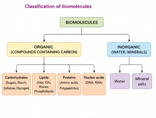
There is a wide diversity in living organisms in our biosphere. Are all living organisms made of the same chemicals, i.e. elements and compounds? You have learnt in chemistry how elemental analysis is performed. If we perform such an analysis on a plant tissue, animal tissue or a microbial paste, we obtain a list of elements like carbon, hydrogen, oxygen and several others, and their respective content per unit mass of living tissue. If the same analysis is performed on a piece of earth's crust as an example of non-living matter, we obtain a similar list. All the elements present in a sample of earth's crust are also present in a sample of living tissue. However, a closer examination reveals that the relative abundance of carbon and hydrogen with respect to other elements is higher in any living organism than in earth's crust. This chapter covers how biomolecules are analysed, the four major classes (carbohydrates, proteins, lipids, nucleic acids), the structure of proteins and polysaccharides, the bonds that link monomers, and enzymes. NEET asks two to three questions every year, mostly on enzymes (classification, inhibition, cofactors), amino acids, protein structure and nucleotides.
- Acid-soluble vs acid-insoluble fractions; why lipids appear in the acid-insoluble fraction.
- Amino acids (zwitterion, essential vs non-essential); fatty acids (palmitic acid has 16 C); lecithin.
- Nucleosides vs nucleotides; purines and pyrimidines.
- Primary to quaternary structure of proteins (haemoglobin: 2α + 2β); peptide, glycosidic and phosphodiester bonds; B-DNA dimensions.
- Polysaccharides: cellulose (homopolymer, no iodine colour), starch (blue-black with iodine), inulin (fructose), chitin.
- Enzymes: active site, activation energy, Km, factors affecting activity, competitive inhibition (malonate–succinic dehydrogenase), six classes, cofactors (prosthetic group, coenzyme, metal ion).
2. How to Analyse Chemical Composition
To find out the inorganic and organic compounds in living organisms, we take a living tissue (a vegetable or a piece of liver) and grind it in trichloroacetic acid (Cl₃CCOOH) using a mortar and a pestle. We obtain a thick slurry. If we strain this through a cheesecloth or cotton, we obtain two fractions. One is called the filtrate or more technically, the acid-soluble pool, and the second, the retentate or the acid-insoluble fraction. Scientists have found thousands of organic compounds in the acid-soluble pool.
In the above analysis, we extract the compounds, then subject the extract to various separation techniques until one has separated a compound from all other compounds. In other words, one isolates and purifies a compound. Analytical techniques, when applied to the compound, give us an idea of the molecular formula and the probable structure of the compound. All the carbon compounds that we get from living tissues can be called biomolecules. However, living organisms have also got inorganic elements and compounds in them. A slightly different but destructive experiment has to be done to find this out. One weighs a small amount of a living tissue (say a leaf or liver; this is called wet weight) and dries it. All the water evaporates. The remaining material gives dry weight. Now if the tissue is fully burnt, all the carbon compounds are oxidised to gaseous form (CO₂, water vapour) and are removed. What is remaining is called the "ash". This ash contains inorganic elements (like calcium, magnesium etc.). Inorganic compounds like sulphate, phosphate etc. are also seen in the acid-soluble fraction.
| Component | % of total cellular mass | NEET Note |
|---|---|---|
| Water | 70–90 | Most abundant chemical in living organisms |
| Proteins | 10–15 | Most abundant organic compound in animal cells |
| Carbohydrates | 3 | — |
| Lipids | 2 | — |
| Nucleic acids | 5–7 | — |
| Ions | 1 | — |
3. Small Biomolecules (Acid-soluble Pool)
Amino acids
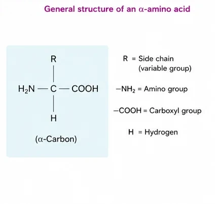
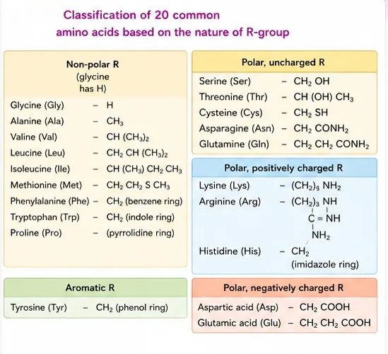
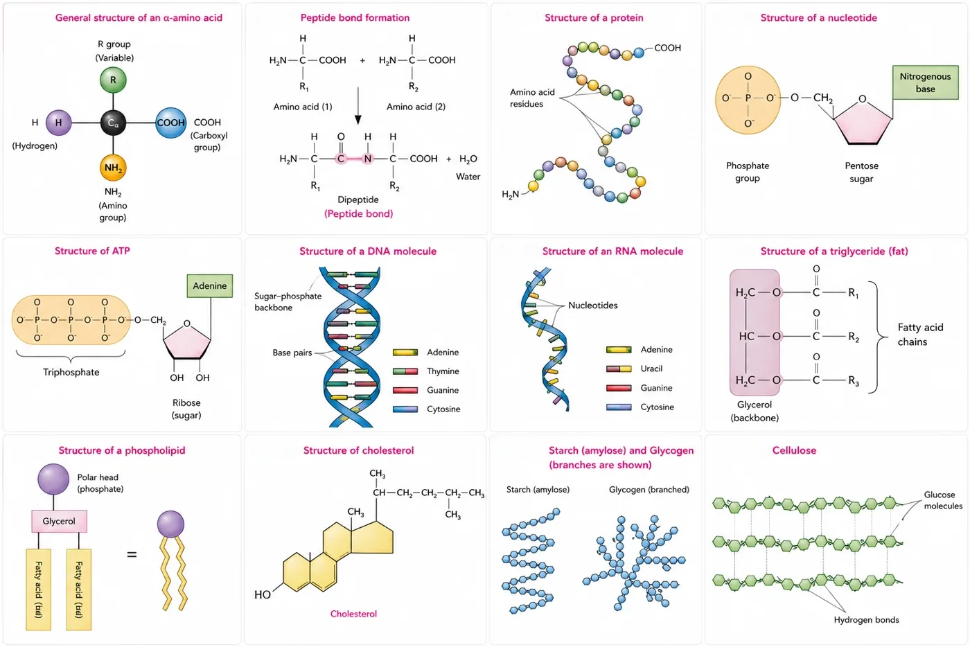
Amino acids are organic compounds containing an amino group and an acidic group as substituents on the same carbon, i.e. the α-carbon. Hence, they are called α-amino acids. They are substituted methanes. There are four substituent groups occupying the four valency positions. These are hydrogen, carboxyl group, amino group and a variable group designated as the R group. Based on the nature of the R group there are many amino acids. However, those which occur in proteins are only of twenty types. The R group in these proteinaceous amino acids could be a hydrogen (the amino acid is called glycine), a methyl group (alanine), hydroxy methyl (serine), etc.
The chemical and physical properties of amino acids are essentially of the amino, carboxyl and the R functional groups. Based on the number of amino and carboxyl groups, there are acidic (e.g. glutamic acid), basic (lysine) and neutral (valine) amino acids. Similarly, there are aromatic amino acids (tyrosine, phenylalanine, tryptophan). A particular property of amino acids is the ionisable nature of –NH₂ and –COOH groups. Hence, in solutions of different pHs, the structure of amino acids changes. The form in which both groups are ionised (–NH₃⁺ and –COO⁻) is called a zwitterionic form.
Lipids
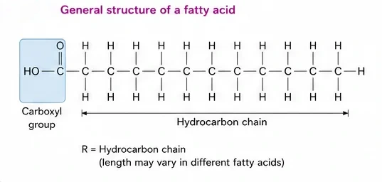
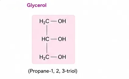
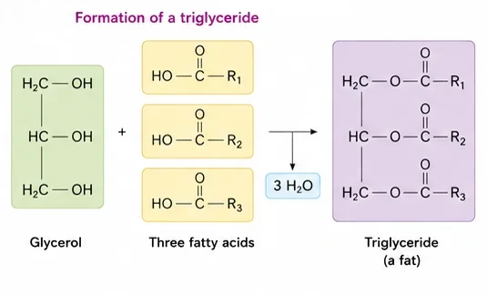
Lipids are generally water insoluble. They could be simple fatty acids. A fatty acid has a carboxyl group attached to an R group. The R group could be a methyl (–CH₃), or ethyl (–C₂H₅), or a higher number of –CH₂ groups (1 carbon to 19 carbons). For example, palmitic acid has 16 carbons including the carboxyl carbon. Arachidonic acid has 20 carbon atoms including the carboxyl carbon. Fatty acids could be saturated (without double bond) or unsaturated (with one or more C=C double bonds). Another simple lipid is glycerol, which is trihydroxy propane. Many lipids have both glycerol and fatty acids. Here the fatty acids are found esterified with glycerol. They can be then monoglycerides, diglycerides and triglycerides. These are also called fats and oils based on melting point. Oils have lower melting point (e.g. gingelly oil) and hence remain as oil in winters. Some lipids have phosphorous and a phosphorylated organic compound in them. These are phospholipids. They are found in cell membranes. Lecithin is one example. Some tissues, especially the neural tissues, have lipids with more complex structures.
Nitrogen bases, nucleosides and nucleotides
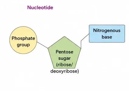
Living organisms have a number of carbon compounds in which heterocyclic rings can be found. Some of these are nitrogen bases — adenine, guanine, cytosine, uracil and thymine. When found attached to a sugar, they are called nucleosides. If a phosphate group is also found esterified to the sugar, they are called nucleotides. Adenosine, guanosine, thymidine, uridine and cytidine are nucleosides. Adenylic acid, thymidylic acid, guanylic acid, uridylic acid and cytidylic acid are nucleotides. Nucleic acids like DNA and RNA consist of nucleotides only. DNA and RNA function as genetic material.
| Category | Members | Ring | NEET Note |
|---|---|---|---|
| Purines | Adenine, guanine | Double ring | Present in both DNA and RNA |
| Pyrimidines | Cytosine, thymine, uracil | Single ring | Thymine only in DNA; uracil only in RNA |
| Nucleoside | Base + sugar | — | Adenosine, cytidine, uridine |
| Nucleotide | Base + sugar + phosphate | — | Adenylic acid, uridylic acid |
Primary and secondary metabolites
The most exciting aspect of chemistry deals with isolating thousands of compounds, small and big, from living organisms, determining their structure and, if possible, synthesising them. Compounds found in animal tissues, like amino acids, sugars and fatty acids, have identifiable functions in normal physiological processes; these are called primary metabolites. However, when one analyses plant, fungal and microbial cells, one would see thousands of compounds other than these, called secondary metabolites. While primary metabolites have identifiable functions and play known roles in normal physiological processes, we do not at the moment understand the role or functions of all the secondary metabolites in host organisms. However, many of them are useful to "human welfare" (e.g. rubber, drugs, spices, scents and pigments). Some secondary metabolites have ecological importance.
| Category | Examples | NEET Note |
|---|---|---|
| Pigments | Carotenoids, anthocyanins | — |
| Alkaloids | Morphine, codeine | — |
| Terpenoids | Monoterpenes, diterpenes | — |
| Essential oils | Lemon grass oil | — |
| Toxins | Abrin, ricin | Ricin from castor seeds |
| Lectins | Concanavalin A | — |
| Drugs | Vinblastin, curcumin | Vinblastin is anticancer |
| Polymeric substances | Rubber, gums, cellulose | — |
4. Biomacromolecules
There is one feature common to all those compounds found in the acid-soluble pool. They have molecular weights ranging from 18 to around 800 daltons (Da) approximately. The acid-insoluble fraction has only four types of organic compounds, i.e. proteins, nucleic acids, polysaccharides and lipids. These classes of compounds, with the exception of lipids, have molecular weights in the range of ten thousand daltons and above. For this very reason, biomolecules, i.e. chemical compounds found in living organisms, are of two types. One, those which have molecular weights less than one thousand dalton and are usually referred to as micromolecules or simply biomolecules, while those which are found in the acid-insoluble fraction are called macromolecules or biomacromolecules.
Lipids have molecular weights that do not exceed 800 Da, yet they come in the acid-insoluble fraction. The reason: lipids are arranged into structures like the cell membrane and other membranes. When we grind a tissue, we are disrupting the cell structure. Cell membranes and other membranes are broken into pieces and form vesicles which are not water soluble. These membrane fragments are separated along with the acid-insoluble pool. Hence lipids are not strictly macromolecules.
5. Proteins
Proteins are polypeptides. They are linear chains of amino acids linked by peptide bonds. Each protein is a polymer of amino acids. As there are 20 types of amino acids (e.g. alanine, cysteine, proline, tryptophan, lysine etc.), a protein is a heteropolymer and not a homopolymer. A homopolymer has only one type of monomer repeating "n" number of times. This information about the amino acid content is important, as later in your nutrition lessons you will learn that certain amino acids are essential for our health and they have to be supplied through our diet. Hence, dietary proteins are the source of essential amino acids. Therefore, amino acids can be essential or non-essential. The latter are those which our body can make, while we get essential amino acids through our diet/food.
Proteins carry out many functions in living organisms; some transport nutrients across the cell membrane, some fight infectious organisms, some are hormones, some are enzymes etc. Collagen is the most abundant protein in the animal world, and Ribulose bisphosphate Carboxylase-Oxygenase (RuBisCO) is the most abundant protein in the whole of the biosphere.
| Protein | Function | NEET Note |
|---|---|---|
| Collagen | Intercellular ground substance | Most abundant animal protein |
| Trypsin | Enzyme | — |
| Insulin | Hormone | — |
| Antibody | Fights infectious agents | — |
| Receptor | Sensory reception (smell, taste, hormone etc.) | — |
| GLUT-4 | Enables glucose transport into cells | — |
Peptide bond
In a polypeptide or a protein, amino acids are linked by a peptide bond, which is formed when the carboxyl (–COOH) group of one amino acid reacts with the amino (–NH₂) group of the next amino acid with the elimination of a water moiety (the process is called dehydration).
Structure of proteins
- Primary structure: the positional information in a protein — which is the first amino acid, which is second, and so on. A protein is imagined as a line, the left end represented by the first amino acid (the N-terminal amino acid) and the right end represented by the last amino acid (the C-terminal amino acid).
- Secondary structure: a protein thread does not exist throughout as an extended rigid rod. The thread is folded in the form of a helix (similar to a revolving staircase). Of course, only some portions of the protein thread are arranged in the form of a helix. In proteins, only right-handed helices are observed. Other regions of the protein thread are folded into other forms.
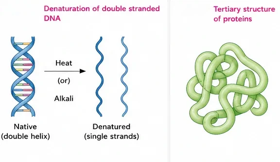
- Tertiary structure: the long protein chain is also folded upon itself like a hollow woollen ball. This gives us a 3-dimensional view of a protein. Tertiary structure is absolutely necessary for the many biological activities of proteins.
- Quaternary structure: some proteins are an assembly of more than one polypeptide or subunits. The manner in which these individual folded polypeptides or subunits are arranged with respect to each other (e.g. linear string of spheres, spheres arranged one upon each other in the form of a cube or plate etc.) is the architecture of a protein, otherwise called the quaternary structure. Adult human haemoglobin consists of 4 subunits. Two of these are identical to each other. Hence, two subunits of α type and two subunits of β type together constitute the human haemoglobin (Hb).
6. Polysaccharides
The acid-insoluble pellet also has polysaccharides (carbohydrates) as another class of macromolecules. Polysaccharides are long chains of sugars. They are threads (literally a cotton thread) containing different monosaccharides as building blocks.
- Cellulose is a polymeric polysaccharide consisting of only one type of monosaccharide, i.e. glucose. Cellulose is a homopolymer.
- Starch is a variant of this but present as a store-house of energy in plant tissues.
- Animals have another variant called glycogen.
- Inulin is a polymer of fructose.
- In a polysaccharide chain (say glycogen), the right end is called the reducing end and the left end is called the non-reducing end. It has branches, as shown in the form of a cartoon.
- Starch forms helical secondary structures. In fact, starch can hold I₂ molecules in the helical portion. The starch–I₂ is blue in colour. Cellulose does not contain complex helices and hence cannot hold I₂.
- Plant cell walls are made of cellulose. Paper made from plant pulp and cotton fibre is cellulosic.
- There are more complex polysaccharides in nature. They have as building blocks amino-sugars and chemically modified sugars (e.g. glucosamine, N-acetyl galactosamine etc.). Exoskeletons of arthropods, for example, have a complex polysaccharide called chitin. These complex polysaccharides are mostly homopolymers.
| Polysaccharide | Monomer | Found in | NEET Note |
|---|---|---|---|
| Starch | Glucose | Plant storage | Helical; blue with iodine |
| Glycogen | Glucose | Animal storage (liver, muscle) | Highly branched |
| Cellulose | Glucose | Plant cell walls, paper, cotton | No helix; no colour with iodine |
| Inulin | Fructose | Dahlia tubers | Used to measure GFR |
| Chitin | Amino-sugars (N-acetyl glucosamine) | Arthropod exoskeleton, fungal walls | Complex homopolymer |
7. Nucleic Acids
The other type of macromolecule that one would find in the acid-insoluble fraction of any living tissue is the nucleic acid. These are polynucleotides. Together with polysaccharides and polypeptides, these comprise the true macromolecular fraction of any living tissue or cell. For nucleic acids, the building block is a nucleotide. A nucleotide has three chemically distinct components. One is a heterocyclic compound, the second is a monosaccharide and the third a phosphoric acid or phosphate.
- The heterocyclic compounds in nucleic acids are the nitrogenous bases named adenine, guanine, uracil, cytosine and thymine. Adenine and guanine are substituted purines, while the rest are substituted pyrimidines. The skeletal heterocyclic ring is called purine and pyrimidine respectively.
- The sugar found in polynucleotides is either ribose (a monosaccharide pentose) or 2'-deoxyribose. A nucleic acid containing deoxyribose is called deoxyribonucleic acid (DNA), while that which contains ribose is called ribonucleic acid (RNA).
8. Nature of Bond Linking Monomers in a Polymer
| Polymer | Bond | Formed between | NEET Note |
|---|---|---|---|
| Polypeptide / protein | Peptide bond | Carboxyl group of one amino acid and amino group of the next, with loss of water | Dehydration |
| Polysaccharide | Glycosidic bond | Two carbon atoms of two adjacent monosaccharides, by dehydration | — |
| Nucleic acid | Phosphodiester bond | Phosphate links the 3'-carbon of one sugar of one nucleotide to the 5'-carbon of the sugar of the succeeding nucleotide | The bond between phosphate and hydroxyl group of sugar is an ester bond; there is one such ester bond on either side, hence phosphodiester |
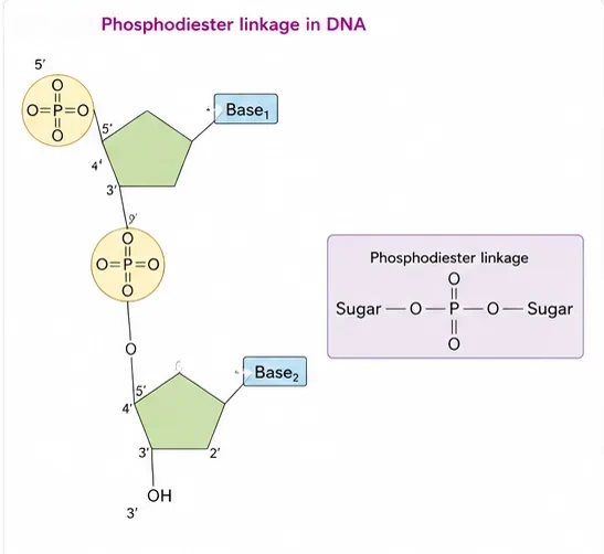
Nucleic acids exhibit a wide variety of secondary structures. For example, one of the secondary structures exhibited by DNA is the famous Watson–Crick model. This model says that DNA exists as a double helix. The two strands of polynucleotides are antiparallel, i.e. run in the opposite direction. The backbone is formed by the sugar–phosphate–sugar chain. The nitrogen bases are projected more or less perpendicular to this backbone but face inside. A and G of one strand compulsorily base pair with T and C, respectively, on the other strand. There are two hydrogen bonds between A and T and three hydrogen bonds between G and C. Each strand appears like a helical staircase. Each step of ascent is represented by a pair of bases. At each step of ascent, the strand turns 36°. One full turn of the helical strand would involve ten steps or ten base pairs. The pitch would be 34 Å. The rise per base pair would be 3.4 Å. This form of DNA with the above-mentioned salient features is called B-DNA.
10 bp per turn · pitch 34 Å (3.4 nm) · rise 3.4 Å per bp · 36° turn per step · A=T (2 H-bonds), G≡C (3 H-bonds) · antiparallel strands · right-handed. These numbers return in Class 12 (Molecular Basis of Inheritance).
9. Dynamic State of Body Constituents — Concept of Metabolism
What we have learnt till now is that living organisms, be it a simple bacterial cell, a protozoan, a plant or an animal, contain thousands of organic compounds. These compounds or biomolecules are present in certain concentrations. One of the greatest discoveries ever made was the observation that all these biomolecules have a turnover. This means that they are constantly being changed into some other biomolecules and also made from some other biomolecules. This breaking and making is through chemical reactions constantly occurring in living organisms. Together all these chemical reactions are called metabolism. Each of the metabolic reactions results in the transformation of biomolecules. A few examples of such metabolic transformations are: removal of CO₂ from amino acids making an amino acid into an amine, removal of amino group in a nucleotide base, hydrolysis of a glycosidic bond in a disaccharide, etc. The majority of these metabolic reactions do not occur in isolation but are always linked to some other reactions. In other words, metabolites are converted into each other in a series of linked reactions called metabolic pathways. Another feature of these metabolic reactions is that every chemical reaction is a catalysed reaction. There is no uncatalysed metabolic conversion in living systems. The catalysts which hasten the rate of a given metabolic conversion are also proteins. These proteins with catalytic power are named enzymes.
10. Enzymes
Almost all enzymes are proteins. There are some nucleic acids that behave like enzymes. These are called ribozymes. One can depict an enzyme by a line diagram. An enzyme, like any protein, has a primary structure, i.e. amino acid sequence of the protein. An enzyme, like any protein, has secondary and tertiary structure. When you look at a tertiary structure, you will notice that the backbone of the protein chain folds upon itself, the chain criss-crosses itself and hence many crevices or pockets are made. One such pocket is the "active site". An active site of an enzyme is a crevice or pocket into which the substrate fits. Thus enzymes, through their active site, catalyse reactions at a high rate.
Enzyme catalysts differ from inorganic catalysts in many ways, but one major difference needs mention. Inorganic catalysts work efficiently at high temperatures and high pressures, while enzymes get damaged at high temperatures (say above 40°C). However, enzymes isolated from organisms who normally live under extremely high temperatures (e.g. hot vents and sulphur springs) are stable and retain their catalytic power even at high temperatures (up to 80°–90°C). Thermal stability is thus an important quality of such enzymes isolated from thermophilic organisms.
How do enzymes bring about such high rates?
Chemical compounds undergo two types of changes. A physical change simply refers to a change in shape without breaking of bonds. Another physical process is a change in the state of matter: when ice melts into water, or when water becomes vapour. However, when bonds are broken and new bonds are formed during transformation, this is called a chemical reaction. The rate of a physical or chemical process refers to the amount of product formed per unit time. Rate can also be called velocity if the direction is specified. Rates of physical and chemical processes are influenced by temperature among other factors. A general rule of thumb is that the rate doubles or decreases by half for every 10°C change in either direction. Catalysed reactions proceed at rates vastly higher than that of uncatalysed ones. For example, CO₂ + H₂O ⇌ H₂CO₃: in the absence of any enzyme this reaction is very slow, with about 200 molecules of H₂CO₃ being formed in an hour. However, by using the enzyme present within the cytoplasm called carbonic anhydrase, the reaction speeds dramatically, with about 600,000 molecules being formed every second. The enzyme has accelerated the reaction rate by about 10 million times. The power of enzymes is incredible indeed!
In any reaction, the substrate (S) has to bind the enzyme at its active site within a given cleft or pocket. The substrate has to diffuse towards the active site. There is thus an obligatory formation of an "ES" complex. E stands for enzyme. This complex formation is a transient phenomenon. During the state where substrate is bound to the enzyme active site, a new structure of the substrate called the transition state structure is formed. Very soon, after the expected bond breaking/making is completed, the product is released from the active site. In other words, the structure of the substrate gets transformed into the structure of the product(s). The pathway of this transformation must go through the so-called transition state structure.
All other intermediate structural states are unstable. Stability is something related to energy status of the molecule or the structure. If "P" is at a lower level than "S", the reaction is an exothermic reaction. One need not supply energy (by heating) in order to form the product. However, whether it is an exothermic or spontaneous reaction or an endothermic or energy-requiring reaction, the "S" has to go through a much higher energy state or transition state. The difference in average energy content of "S" from that of this transition state is called activation energy. Enzymes eventually bring down this energy barrier, making the transition of "S" to "P" more easy.
Nature of enzyme action
Each enzyme (E) has a substrate (S)-binding site in its molecule so that a highly reactive enzyme–substrate complex (ES) is produced. This complex is short-lived and dissociates into its product(s) P and the unchanged enzyme, with an intermediate formation of the enzyme–product complex (EP). The formation of the ES complex is essential for catalysis: E + S ⇌ ES → EP → E + P. The catalytic cycle of an enzyme action can be described in the following steps:
- First, the substrate binds to the active site of the enzyme, fitting into the active site.
- The binding of the substrate induces the enzyme to alter its shape, fitting more tightly around the substrate.
- The active site of the enzyme, now in close proximity of the substrate, breaks the chemical bonds of the substrate and the new enzyme–product complex is formed.
- The enzyme releases the products of the reaction and the free enzyme is ready to bind to another molecule of the substrate and run through the catalytic cycle once again.
Factors affecting enzyme activity
- Temperature and pH: enzymes generally function in a narrow range of temperature and pH. Each enzyme shows its highest activity at a particular temperature and pH called the optimum temperature and optimum pH. Activity declines both below and above the optimum value. Low temperature preserves the enzyme in a temporarily inactive state, whereas high temperature destroys enzymatic activity because proteins are denatured by heat.
- Concentration of substrate: with the increase in substrate concentration, the velocity of the enzymatic reaction rises at first. The reaction ultimately reaches a maximum velocity (Vmax), which is not exceeded by any further rise in concentration of the substrate. This is because the enzyme molecules are fewer than the substrate molecules, and after saturation of these molecules, there are no free enzyme molecules to bind with the additional substrate molecules.
- The activity of an enzyme is also sensitive to the presence of specific chemicals that bind to the enzyme. When the binding of the chemical shuts off enzyme activity, the process is called inhibition and the chemical is called an inhibitor. When the inhibitor closely resembles the substrate in its molecular structure and inhibits the activity of the enzyme, it is known as a competitive inhibitor. Due to its close structural similarity with the substrate, the inhibitor competes with the substrate for the substrate-binding site of the enzyme. Consequently, the substrate cannot bind and, as a result, the enzyme action declines, e.g. inhibition of succinic dehydrogenase by malonate, which closely resembles the substrate succinate in structure. Such competitive inhibitors are often used in the control of bacterial pathogens.
Classification and nomenclature of enzymes
Thousands of enzymes have been discovered, isolated and studied. Most of these enzymes have been classified into different groups based on the type of reactions they catalyse. Enzymes are divided into 6 classes, each with 4–13 subclasses, and named accordingly by a four-digit number.
| Class | Reaction catalysed | NEET Note |
|---|---|---|
| 1. Oxidoreductases / dehydrogenases | Oxidoreduction between two substrates S and S': S reduced + S' oxidised → S oxidised + S' reduced | e.g. succinic dehydrogenase |
| 2. Transferases | Transfer of a group G (other than hydrogen) between a pair of substrates S and S': S–G + S' → S + S'–G | e.g. kinases |
| 3. Hydrolases | Hydrolysis of ester, ether, peptide, glycosidic, C–C, C–halide or P–N bonds | Digestive enzymes |
| 4. Lyases | Removal of groups from substrates by mechanisms other than hydrolysis, leaving double bonds | e.g. aldolase |
| 5. Isomerases | Inter-conversion of optical, geometric or positional isomers | — |
| 6. Ligases | Linking together of 2 compounds, e.g. joining C–O, C–S, C–N, P–O etc. bonds | e.g. DNA ligase |
Co-factors
Enzymes are composed of one or several polypeptide chains. However, there are a number of cases in which non-protein constituents called co-factors are bound to the enzyme to make the enzyme catalytically active. In these instances, the protein portion of the enzyme is called the apoenzyme. Three kinds of co-factors may be identified: prosthetic groups, co-enzymes and metal ions.
- Prosthetic groups are organic compounds and are distinguished from other co-factors in that they are tightly bound to the apoenzyme. For example, in peroxidase and catalase, which catalyse the breakdown of hydrogen peroxide to water and oxygen, haem is the prosthetic group and it is a part of the active site of the enzyme.
- Co-enzymes are also organic compounds but their association with the apoenzyme is only transient, usually occurring during the course of catalysis. Furthermore, co-enzymes serve as co-factors in a number of different enzyme-catalysed reactions. The essential chemical components of many coenzymes are vitamins, e.g. coenzymes nicotinamide adenine dinucleotide (NAD) and NADP contain the vitamin niacin.
- A number of enzymes require metal ions for their activity, which form coordination bonds with side chains at the active site and at the same time form one or more coordination bonds with the substrate, e.g. zinc is a cofactor for the proteolytic enzyme carboxypeptidase.
Catalytic activity is lost when the co-factor is removed from the enzyme, which testifies that they play a crucial role in the catalytic activity of the enzyme.
- Competitive inhibition: malonate inhibits succinic dehydrogenase (resembles succinate).
- Prosthetic group: haem in peroxidase and catalase. Coenzyme: NAD/NADP contain niacin. Metal ion: Zn²⁺ for carboxypeptidase.
- Carbonic anhydrase speeds its reaction ~10 million times.
- Enzymes lower activation energy; they do not change the equilibrium.
- Ribozymes are RNA enzymes (e.g. peptidyl transferase activity of rRNA).
11. Master Summary Table
| Biomolecule | Monomer / unit | Bond | NEET Note |
|---|---|---|---|
| Protein | 20 amino acids | Peptide | Heteropolymer; Hb 2α + 2β; RuBisCO most abundant |
| Polysaccharide | Monosaccharides | Glycosidic | Cellulose homopolymer; starch–I₂ blue |
| Nucleic acid | Nucleotides | Phosphodiester (3'–5') | B-DNA: 10 bp/turn, 34 Å pitch |
| Lipid | Fatty acids + glycerol | Ester | Not a true macromolecule; < 800 Da |
| Enzyme | Protein (or RNA ribozyme) | — | 6 classes; cofactors; Km, Vmax |
12. Mind Map
- Acid-soluble pool
- Acid-insoluble fraction
- Ash (inorganic)
- Amino acids, sugars
- Fatty acids, glycerol
- Nucleotides; secondary metabolites
- Proteins (4 levels)
- Polysaccharides
- Nucleic acids (B-DNA)
- Active site, Ea
- Factors, inhibition
- 6 classes, cofactors
13. Tips and Tricks
- Enzyme classes in order: "Over The Hill Lives Indian Lion" — Oxidoreductase, Transferase, Hydrolase, Lyase, Isomerase, Ligase.
- Purines: "Pure As Gold" — Adenine, Guanine. Pyrimidines: "CUT the py" — Cytosine, Uracil, Thymine.
- Bonds: Protein → Peptide; Sugar → glycosidic; Nucleic acid → phosphodiester.
- Cofactors: Prosthetic = Permanently (tightly) bound; Coenzyme = Casual (transient).
- Calling lipids true macromolecules (they come in the acid-insoluble fraction only because of membrane vesicles).
- Saying cellulose gives a blue colour with iodine (only starch does).
- Saying proteins are homopolymers (they are heteropolymers).
- Saying enzymes change the equilibrium of a reaction (they only speed up reaching it).
- Confusing nucleoside (no phosphate) with nucleotide (with phosphate).
14. Important One-liners
- Trichloroacetic acid is used to separate the acid-soluble and acid-insoluble fractions.
- Water forms 70–90% of total cellular mass.
- Glycine is the simplest amino acid (R = H).
- Palmitic acid has 16 carbon atoms; arachidonic acid has 20.
- Lecithin is a phospholipid.
- Ricin and abrin are toxins; vinblastin is an anticancer drug.
- Collagen is the most abundant protein in animals; RuBisCO in the biosphere.
- Only right-handed helices are found in proteins.
- Inulin is a polymer of fructose.
- Chitin forms the exoskeleton of arthropods.
- A–T pairs have two hydrogen bonds; G–C pairs have three.
- Haem is the prosthetic group of catalase and peroxidase.
15. NEET-pattern Questions with Detailed Answers
These questions are original and based on concepts that NEET UG asks again and again.
The most abundant protein in the whole biosphere is:
- Collagen
- Haemoglobin
- RuBisCO
- Insulin
Show solution
RuBisCO is the most abundant protein in the biosphere because every photosynthetic organism makes large amounts of it. Collagen is the most abundant protein in animals only.
Answer: (C)
Which of the following is a purine?
- Cytosine
- Thymine
- Uracil
- Guanine
Show solution
Adenine and guanine are purines (double ring). Cytosine, thymine and uracil are pyrimidines (single ring).
Answer: (D)
Lipids appear in the acid-insoluble fraction because:
- Their molecular weight exceeds 10,000 Da
- They form insoluble membrane vesicles when tissue is ground
- They are polymers of fatty acids
- They are bound to nucleic acids
Show solution
Lipids are small (under 800 Da). When the tissue is ground, the membranes they form break into water-insoluble vesicles, which separate with the acid-insoluble pellet.
Answer: (B)
Malonate inhibits succinic dehydrogenase because it:
- Denatures the enzyme
- Resembles succinate and competes for the active site
- Binds permanently to an allosteric site
- Removes the metal cofactor
Show solution
Malonate is structurally similar to succinate, the substrate. It competes for the substrate-binding site, which is competitive inhibition.
Answer: (B)
In B-DNA, the pitch of the helix and the rise per base pair are:
- 34 Å and 3.4 Å
- 3.4 Å and 34 Å
- 20 Å and 3.4 Å
- 34 Å and 0.34 Å
Show solution
One turn of B-DNA has 10 base pairs, with a pitch of 34 Å and a rise of 3.4 Å per base pair. Option (B) reverses the values.
Answer: (A)
The coenzymes NAD and NADP contain the vitamin:
- Riboflavin
- Niacin
- Thiamine
- Folic acid
Show solution
NAD and NADP are built from niacin (nicotinic acid). Riboflavin forms FAD and FMN, and thiamine forms TPP.
Answer: (B)
Assertion (A): Starch gives a blue colour with iodine but cellulose does not.
Reason (R): Starch forms helical secondary structures that hold I₂, while cellulose lacks complex helices.
- Both A and R are true, and R explains A
- Both A and R are true, but R does not explain A
- A is true, R is false
- A is false, R is true
Show solution
Iodine fits inside the helical parts of starch to give the blue colour. Cellulose chains are straight and cannot hold iodine. R explains A.
Answer: (A)
Enzymes that catalyse the removal of groups from substrates by mechanisms other than hydrolysis, leaving double bonds, are:
- Hydrolases
- Lyases
- Ligases
- Transferases
Show solution
Lyases remove groups without hydrolysis and leave double bonds. Hydrolases add water to break bonds, ligases join two molecules, and transferases move groups from one substrate to another.
Answer: (B)
Which statement about co-factors is correct?
- Coenzymes are tightly and permanently bound to the apoenzyme
- Haem is the prosthetic group of catalase and peroxidase
- Zinc is the coenzyme of carboxypeptidase
- Removing a cofactor does not affect catalytic activity
Show solution
Haem is the tightly bound prosthetic group of catalase and peroxidase. Coenzymes bind only for a short time, zinc is a metal-ion cofactor (not a coenzyme), and removing a cofactor destroys activity.
Answer: (B)
Match: (a) Peptide bond (b) Glycosidic bond (c) Phosphodiester bond (d) Hydrogen bond — (i) Between complementary bases of DNA (ii) Between monosaccharides (iii) Between amino acids (iv) Between sugars of adjacent nucleotides through phosphate.
- a-iii, b-ii, c-iv, d-i
- a-ii, b-iii, c-iv, d-i
- a-iii, b-iv, c-ii, d-i
- a-iii, b-ii, c-i, d-iv
Show solution
Peptide bonds join amino acids, glycosidic bonds join sugars, phosphodiester bonds join nucleotides through a phosphate, and hydrogen bonds hold complementary DNA bases together.
Answer: (A)
16. Quick Revision Summary
- Grinding in trichloroacetic acid gives an acid-soluble pool (micromolecules, 18–800 Da) and an acid-insoluble fraction (proteins, nucleic acids, polysaccharides, lipids).
- Amino acids: α-carbon with H, NH₂, COOH, R; 20 in proteins; zwitterion; essential vs non-essential.
- Lipids: fatty acids (palmitic 16 C, arachidonic 20 C), glycerol, triglycerides, phospholipids (lecithin).
- Nucleotides = base + sugar + phosphate; purines A, G; pyrimidines C, T, U.
- Proteins: heteropolymers; primary → quaternary (Hb 2α + 2β); collagen and RuBisCO. Polysaccharides: cellulose, starch, glycogen, inulin, chitin.
- Bonds: peptide, glycosidic, phosphodiester. B-DNA: antiparallel, 10 bp per turn, 34 Å pitch, 3.4 Å rise.
- Enzymes: active site, ES complex, lower activation energy; affected by temperature, pH, substrate concentration (Vmax, Km) and inhibitors; 6 classes; cofactors — prosthetic group (haem), coenzyme (NAD, niacin), metal ion (Zn²⁺).