
1. Chapter at a Glance
All organisms, even the largest, start their life from a single cell. Growth and reproduction depend on cells dividing: one parent cell forms two or more daughter cells. The sequence of events by which a cell duplicates its genome, synthesises its other constituents and eventually divides into two daughter cells is called the cell cycle. This chapter explains the phases of the cell cycle, the two kinds of cell division — mitosis (equational division) and meiosis (reduction division) — and their significance. A short bridge section shows what happens to DNA inside the dividing cell, linking to Class 12 molecular biology. NEET asks about two questions from this chapter, usually on the events of each phase, prophase I sub-stages and the changes in DNA content and chromosome number.
- Phases of the cell cycle; duration in human cells and yeast; G₀ (quiescent) stage.
- DNA content (C) and chromosome number (n) in G₁, S, G₂ and M.
- Events of prophase, metaphase, anaphase and telophase; cytokinesis in plants vs animals.
- Five sub-stages of prophase I (leptotene, zygotene, pachytene, diplotene, diakinesis).
- Synaptonemal complex, chiasmata, crossing over and the significance of mitosis and meiosis.
2. The Cell Cycle
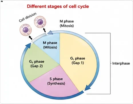
Cell division is a very important process in all living organisms. During division, DNA replication and cell growth also take place. All these processes — cell division, DNA replication and cell growth — must happen in a coordinated way to ensure correct division and the formation of progeny cells with intact genomes. Although cell growth (in terms of cytoplasmic increase) is a continuous process, DNA synthesis occurs only during one specific stage of the cycle. The replicated chromosomes are then distributed to daughter nuclei by a complex series of events, all under genetic control.
- A typical eukaryotic cell, e.g. a human cell in culture, divides about once every 24 hours. The duration varies between organisms and cell types: yeast can complete the cycle in about 90 minutes.
- The cycle has two basic phases: interphase (the period between two successive M phases) and M phase (mitosis phase, when actual division occurs).
- In a 24-hour human cell cycle, cell division proper lasts only about an hour. Interphase lasts more than 95% of the duration of the cycle.
- Interphase is not a resting phase: the cell is preparing for division by undergoing both cell growth and DNA replication in an orderly manner.
| Phase | What happens | NEET Note |
|---|---|---|
| G₁ (Gap 1) | Interval between mitosis and the start of DNA replication. The cell is metabolically active and grows continuously but does not replicate its DNA. | Longest phase in most cells |
| S (Synthesis) | DNA synthesis or replication. DNA per cell doubles: if initial amount is 2C, it becomes 4C. The number of chromosomes does not change (2n stays 2n). | In animal cells, the centriole duplicates in the cytoplasm |
| G₂ (Gap 2) | Proteins are synthesised in preparation for mitosis while cell growth continues. | — |
| G₀ (quiescent stage) | Some cells in adult animals do not appear to divide (e.g. heart cells), and many others divide only occasionally to replace lost or dead cells. Cells that do not divide further exit G₁ to enter G₀. | Cells remain metabolically active but no longer proliferate, unless called upon |
| M (Mitosis) | Karyokinesis (nuclear division) followed by cytokinesis (division of cytoplasm). | Starts with nuclear division |
In animals, mitotic division is seen only in the diploid somatic cells. Plants show mitotic divisions in both haploid and diploid cells. Can you think of examples? (Haploid gametophytes of mosses and ferns divide by mitosis.)
3. M Phase: Mitosis
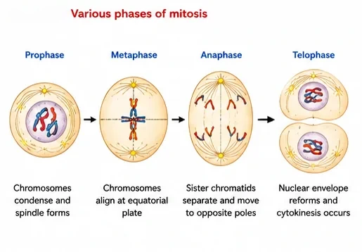
Mitosis is the most dramatic period of the cell cycle, involving a major reorganisation of virtually all components of the cell. Since the number of chromosomes in the parent and progeny cells is the same, it is called equational division. Mitosis is divided into four stages of nuclear division (karyokinesis). It is a progressive process, and clear-cut lines cannot be drawn between stages.
Prophase
- Follows the S and G₂ phases. The new DNA molecules formed are not distinct but intertwined.
- Initiation of condensation of chromosomal material: chromosomes become untangled and compact. Each chromosome now consists of two chromatids attached at the centromere.
- The centrosome (which had duplicated during S) begins to move towards opposite poles. Each centrosome radiates microtubules called asters. The two asters with spindle fibres form the mitotic apparatus.
- At the end of prophase, cells do not show Golgi complexes, endoplasmic reticulum, nucleolus or the nuclear envelope.
Metaphase
- Marked by the complete disintegration of the nuclear envelope; chromosomes are spread through the cytoplasm.
- Condensation of chromosomes is complete, and they can be observed clearly under the microscope. This is the stage at which the morphology of chromosomes is most easily studied.
- A metaphase chromosome is made up of two sister chromatids held together by the centromere. Small disc-shaped structures on the surface of the centromere are called kinetochores; they serve as sites of attachment of spindle fibres.
- Chromosomes come to lie at the equator; one chromatid of each chromosome is connected by its kinetochore to spindle fibres from one pole and its sister to fibres from the opposite pole. The plane of alignment is the metaphase plate.
Anaphase
- Each chromosome arranged at the metaphase plate splits simultaneously: the two daughter chromatids, now called chromosomes of the future daughter nuclei, begin to migrate towards opposite poles.
- As each chromosome moves away from the equatorial plate, the centromere leads, and the arms trail behind.
- So anaphase involves: centromeres split and chromatids separate; chromatids move to opposite poles.
Telophase
- Chromosomes that have reached their respective poles decondense and lose their individuality. The individual chromosomes can no longer be seen; chromatin material collects at each pole.
- The nuclear envelope develops around the chromosome clusters at each pole, forming two daughter nuclei.
- The nucleolus, Golgi complex and ER re-form.
Cytokinesis
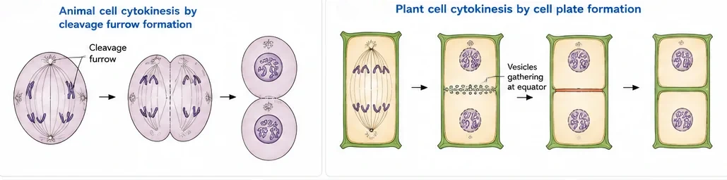
Mitosis divides the duplicated chromosomes into daughter nuclei (karyokinesis); the cell itself divides into two daughter cells by cytokinesis, the division of cytoplasm.
- In animal cells, a furrow appears in the plasma membrane. It gradually deepens and finally joins in the centre, dividing the cytoplasm into two (cleavage furrow).
- Plant cells are enclosed by a relatively rigid cell wall, so the mechanism is different. Wall formation starts in the centre and grows outward to meet the existing lateral walls. It begins with a simple precursor called the cell plate, which represents the middle lamella between the walls of two adjacent cells.
- At the time of cytoplasmic division, organelles such as mitochondria and plastids are distributed between the two daughter cells.
- In some organisms, karyokinesis is not followed by cytokinesis, resulting in a multinucleate condition called a syncytium (e.g. the liquid endosperm of coconut).
Significance of mitosis
- Results in the production of diploid daughter cells with an identical genetic complement (usually).
- Growth of multicellular organisms depends on mitosis.
- Restores the nucleo-cytoplasmic ratio, which is disturbed as a cell grows.
- Cell repair: cells of the upper layer of epidermis, cells of the lining of the gut and blood cells are constantly replaced.
- Mitotic divisions in meristematic tissues — apical and lateral cambium — result in continuous growth of plants throughout their life.
4. Meiosis
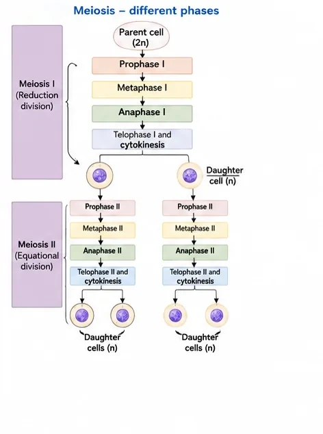
The production of offspring by sexual reproduction includes the fusion of two gametes, each with a complete haploid set of chromosomes. Gametes are formed from specialised diploid cells by meiosis, a specialised kind of cell division that reduces the chromosome number by half, resulting in haploid daughter cells. Meiosis ensures the production of haploid phase in the life cycle of sexually reproducing organisms, whereas fertilisation restores the diploid phase.
- It involves two sequential cycles of nuclear and cell division, meiosis I and meiosis II, but only a single cycle of DNA replication.
- Meiosis I is initiated after the parental chromosomes have replicated in the S phase.
- It involves pairing of homologous chromosomes and recombination between non-sister chromatids.
- Four haploid cells are formed at the end of meiosis II.
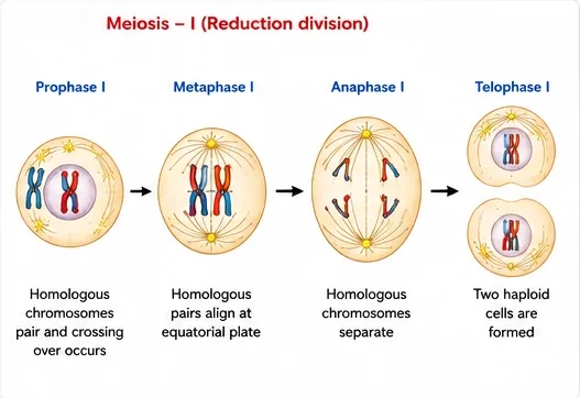
Meiosis I
Prophase I is typically longer and more complex than prophase of mitosis. It is subdivided into five phases based on chromosomal behaviour:
| Sub-stage | Key event | NEET Note |
|---|---|---|
| Leptotene | Chromosomes gradually become visible under the light microscope; compaction continues | "Thin thread" stage |
| Zygotene | Homologous chromosomes start pairing (synapsis), accompanied by formation of the synaptonemal complex. A pair of synapsed homologues is a bivalent or tetrad. | Synapsis begins |
| Pachytene | Bivalents appear clearly as tetrads. Recombination nodules appear, the sites where crossing over occurs between non-sister chromatids of homologous chromosomes. Crossing over is enzyme-mediated (enzyme: recombinase). | Crossing over; longer than leptotene and zygotene |
| Diplotene | The synaptonemal complex dissolves and the homologues of bivalents tend to separate, except at the sites of crossover. These X-shaped structures are called chiasmata. In oocytes of some vertebrates, diplotene can last for months or years. | Chiasmata visible |
| Diakinesis | Terminalisation of chiasmata. Chromosomes are fully condensed and the meiotic spindle is assembled. The nucleolus disappears and the nuclear envelope breaks down. | Transition to metaphase |
- Metaphase I: bivalent chromosomes align on the equatorial plate. Microtubules from opposite poles attach to the kinetochores of homologous chromosomes.
- Anaphase I: homologous chromosomes separate, while sister chromatids remain associated at their centromeres.
- Telophase I: the nuclear membrane and nucleolus reappear; cytokinesis follows. This is called a dyad of cells. In many cases chromosomes do undergo some dispersion, but they do not reach the extremely extended state of interphase.
- The stage between the two meiotic divisions is called interkinesis; it is generally short-lived. There is no replication of DNA during interkinesis.
Meiosis II
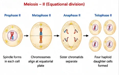
- Initiated immediately after cytokinesis, usually before the chromosomes have fully elongated. It resembles a normal mitosis.
- Prophase II: the nuclear membrane disappears by the end of the stage; chromosomes again become compact.
- Metaphase II: chromosomes align at the equator; microtubules from opposite poles attach to the kinetochores of sister chromatids.
- Anaphase II: simultaneous splitting of the centromere of each chromosome (which was holding the sister chromatids together), allowing them to move to opposite poles.
- Telophase II: the two groups of chromosomes again get enclosed by a nuclear envelope; cytokinesis follows, resulting in a tetrad of cells, i.e. four haploid daughter cells.
Significance of meiosis
- Conserves the specific chromosome number of each species across generations in sexually reproducing organisms, even though, paradoxically, the process itself results in reduction of chromosome number by half.
- Increases genetic variability in the population from one generation to the next (through crossing over and independent assortment). Variations are very important for evolution.
| Feature | Mitosis | Meiosis |
|---|---|---|
| Type of division | Equational | Reductional (I) + equational (II) |
| Number of divisions | One | Two (one DNA replication) |
| Daughter cells | Two, diploid (2n), identical | Four, haploid (n), genetically different |
| Pairing and crossing over | Absent | Present (zygotene and pachytene) |
| Where it occurs | Somatic cells; also haploid cells in plants | Reproductive (germ) cells |
| Anaphase | Sister chromatids separate | Anaphase I: homologues separate; anaphase II: sister chromatids separate |
5. Bridge to Class 12: DNA in the Dividing Cell
The S phase of the cell cycle exists to copy the cell's DNA, so it helps to know how DNA is packed and copied. These topics are studied in detail in Class 12 (Molecular Basis of Inheritance); here is a brief look connected to cell division.
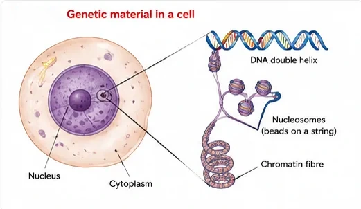
- Packaging: in eukaryotes, DNA is wrapped around positively charged proteins called histones (an octamer of histones) to form a unit called the nucleosome. Nucleosomes look like "beads on a string" under the electron microscope, and they coil further to form chromatin fibres, which condense into chromosomes during prophase.
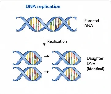
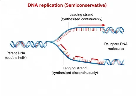
- Replication in the S phase is semiconservative: each new DNA molecule contains one old (parental) strand and one newly made strand. At the replication fork, one strand (the leading strand) is made continuously and the other (the lagging strand) is made in short pieces (Okazaki fragments), because DNA polymerase can work only in the 5′→3′ direction.
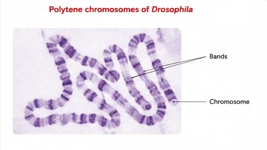
- Polytene chromosomes: in the salivary glands of Drosophila larvae, DNA replicates repeatedly without cell division. The many copies stay side by side, forming giant banded chromosomes — a striking example of DNA replication without division.
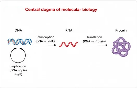
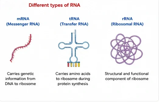
From DNA to protein. The DNA copied during S phase carries genes. A gene's information is copied into RNA (transcription) and then used to make a protein (translation). This flow — DNA → RNA → protein, with DNA also copying itself — is the central dogma of molecular biology, proposed by Francis Crick. Three kinds of RNA take part: mRNA carries the message, tRNA brings amino acids, and rRNA forms the structure of the ribosome.
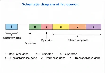
Genes can be switched on and off. Cells do not use all their genes all the time. In bacteria, groups of related genes are controlled together as an operon. The lac operon of E. coli, described by Jacob and Monod, has a regulatory gene (i), a promoter (p), an operator (o) and three structural genes: z (β-galactosidase), y (permease) and a (transacetylase). Lactose acts as the inducer.
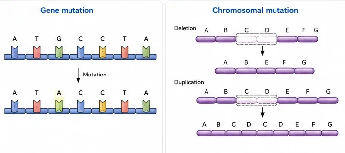
Mistakes in DNA: mutations. Errors during replication or damage can change DNA. A change in a single base pair is a gene (point) mutation; a famous example is sickle-cell anaemia. Changes in the structure of chromosomes — deletion, duplication, inversion or translocation of segments — are chromosomal mutations. Errors in the separation of chromosomes during cell division (non-disjunction) change the chromosome number, as in Down's syndrome (trisomy 21).
6. Master Summary Table
| Stage | Key event | NEET Note |
|---|---|---|
| G₁ | Cell growth | 2C, 2n |
| S | DNA replication; centriole duplicates | 2C → 4C, chromosome number unchanged |
| G₂ | Protein synthesis for mitosis | 4C |
| G₀ | Quiescent, metabolically active | Heart cells, many adult cells |
| Prophase | Chromosome condensation, spindle forms | Nuclear envelope and nucleolus disappear |
| Metaphase | Chromosomes at metaphase plate | Best stage to study chromosome morphology |
| Anaphase | Centromeres split; chromatids move to poles | Centromere leads |
| Telophase | Nuclear envelope re-forms | Chromosomes decondense |
| Pachytene | Crossing over | Recombinase; recombination nodules |
| Diplotene | Chiasmata visible | Synaptonemal complex dissolves |
| Interkinesis | Short gap between meiosis I and II | No DNA replication |
7. Mind Map
- G₁, S, G₂
- M phase
- G₀
- P-M-A-T
- Cytokinesis
- Significance
- Prophase I sub-stages
- Meiosis II
- Variation
- Nucleosome
- Semiconservative replication
- Mutation
8. Tips and Tricks
- Mitosis stages: "Please Meet At Ten" — prophase, metaphase, anaphase, telophase.
- Prophase I: "Lazy Zebras Prefer Deep Dozing" — leptotene, zygotene, pachytene, diplotene, diakinesis.
- "Zygotene = Zipping (synapsis); Pachytene = Partner swap (crossing over); Diplotene = Display of chiasmata; Diakinesis = Disappearance of nucleolus (terminalisation)."
- DNA (C) changes only in S phase (doubles) and at division (halves); chromosome number (n) changes only when centromeres split or homologues separate.
- Saying chromosome number doubles in S phase (only DNA doubles).
- Calling interphase a resting phase.
- Placing crossing over in zygotene (synapsis) instead of pachytene.
- Thinking DNA replicates during interkinesis.
- Saying homologous chromosomes separate in anaphase II (they separate in anaphase I).
9. Important One-liners
- A human cell in culture divides about once in 24 hours; yeast in about 90 minutes.
- Interphase occupies more than 95% of the cell cycle.
- DNA replication occurs in the S phase.
- Cells that stop dividing enter the G₀ stage.
- Metaphase is the best stage to count and study chromosomes.
- Kinetochores are the sites where spindle fibres attach to chromosomes.
- The cell plate in plant cells represents the middle lamella.
- Free-nuclear endosperm of coconut is an example of a syncytium.
- Crossing over occurs in pachytene and is mediated by recombinase.
- Chiasmata become visible in diplotene and are terminalised in diakinesis.
- Meiosis has two divisions but only one round of DNA replication.
10. NEET-pattern Questions with Detailed Answers
These questions are original and based on concepts that NEET UG asks again and again.
DNA replication takes place in which phase of the cell cycle?
- G₁
- S
- G₂
- M
Show solution
The S (synthesis) phase is when DNA doubles from 2C to 4C. G₁ and G₂ are growth and preparation phases, and in the M phase the cell divides.
Answer: (B)
The stage of mitosis best suited for studying the morphology of chromosomes is:
- Prophase
- Metaphase
- Anaphase
- Telophase
Show solution
In metaphase, condensation is complete and the chromosomes lie spread out at the metaphase plate, so each one can be seen clearly. In prophase they are still condensing, and in telophase they decondense again.
Answer: (B)
A cell has 2C DNA and 2n chromosomes in G₁. What will its DNA content and chromosome number be in G₂?
- 4C, 4n
- 4C, 2n
- 2C, 2n
- 2C, 4n
Show solution
In the S phase, DNA doubles to 4C. Each chromosome now has two sister chromatids held at one centromere, so the chromosome number stays 2n. Option (A) wrongly doubles the chromosome number as well.
Answer: (B)
Crossing over between non-sister chromatids of homologous chromosomes takes place during:
- Leptotene
- Zygotene
- Pachytene
- Diakinesis
Show solution
Recombination nodules appear in pachytene, and crossing over happens there, catalysed by recombinase. Synapsis begins in zygotene, chiasmata appear in diplotene, and they are terminalised in diakinesis.
Answer: (C)
In plant cells, cytokinesis takes place by:
- Cleavage furrow from outside
- Cell plate formation from the centre outwards
- Budding
- Constriction of the cell wall
Show solution
The rigid wall prevents a furrow from forming. Instead, vesicles gather at the centre to form a cell plate (the future middle lamella), which grows outwards. A cleavage furrow is the animal-cell method.
Answer: (B)
Which of these is NOT a feature of meiosis?
- Two divisions
- Two rounds of DNA replication
- Pairing of homologous chromosomes
- Four haploid daughter cells
Show solution
Meiosis has two divisions but only one round of DNA replication, which happens in the S phase before meiosis I. There is no replication during interkinesis. The other three options are true features of meiosis.
Answer: (B)
Assertion (A): Heart cells of adult humans are in the G₀ stage.
Reason (R): Cells in G₀ are metabolically inactive.
- Both A and R are true, and R explains A
- Both A and R are true, but R does not explain A
- A is true, R is false
- A is false, R is true
Show solution
Adult heart cells do not divide, so they stay in G₀. However, G₀ cells remain metabolically active; they simply stop proliferating. So A is true and R is false.
Answer: (C)
Match the sub-stages of prophase I with their events: (a) Zygotene (b) Pachytene (c) Diplotene (d) Diakinesis — (i) Terminalisation of chiasmata (ii) Synapsis (iii) Dissolution of synaptonemal complex (iv) Crossing over.
- a-ii, b-iv, c-iii, d-i
- a-iv, b-ii, c-i, d-iii
- a-ii, b-iii, c-iv, d-i
- a-iii, b-iv, c-ii, d-i
Show solution
Synapsis happens in zygotene and crossing over in pachytene. In diplotene the synaptonemal complex dissolves and chiasmata appear. In diakinesis the chiasmata are terminalised.
Answer: (A)
The liquid endosperm of coconut contains thousands of free nuclei. This is because:
- Meiosis is not followed by cytokinesis
- Karyokinesis is not followed by cytokinesis
- Cytokinesis occurs without karyokinesis
- Cells fuse after division
Show solution
Repeated nuclear divisions without division of the cytoplasm produce a multinucleate syncytium. The coconut's liquid endosperm (coconut water) is the classic example. Cytokinesis cannot happen without karyokinesis before it, and the endosperm nuclei divide by mitosis, not meiosis.
Answer: (B)
A diploid organism has 2n = 24. How many chromatids are present in each cell at metaphase I and in each daughter cell at metaphase II?
- 48 and 24
- 24 and 12
- 48 and 48
- 24 and 24
Show solution
After the S phase, each of the 24 chromosomes has 2 chromatids, giving 48 at metaphase I. After meiosis I, each cell has 12 chromosomes, still with 2 chromatids each, giving 24 at metaphase II. The chromatids separate only in anaphase II.
Answer: (A)
11. Quick Revision Summary
- Cell cycle = interphase (G₁, S, G₂; >95% of time) + M phase; human cell 24 h, yeast 90 min; G₀ for non-dividing cells.
- S phase doubles DNA (2C → 4C), not chromosome number.
- Mitosis: prophase (condensation), metaphase (plate, kinetochores), anaphase (centromeres split), telophase (nuclei re-form); cytokinesis by furrow (animals) or cell plate (plants).
- Meiosis: 2 divisions, 1 replication. Prophase I: leptotene, zygotene (synapsis), pachytene (crossing over), diplotene (chiasmata), diakinesis (terminalisation).
- Anaphase I separates homologues; anaphase II separates sister chromatids; four haploid cells.
- Mitosis gives growth and repair; meiosis keeps chromosome number constant and creates variation.