
1. Chapter at a Glance
The neural system provides a point-to-point rapid coordination among organs. This neural coordination is fast but short-lived. As the nerve fibres do not innervate all cells of the body, and the cellular functions need to be continuously regulated, a special kind of coordination and integration has to be provided. This function is carried out by hormones. The neural system and the endocrine system jointly coordinate and regulate the physiological functions in the body. This chapter covers endocrine glands and hormones, the human endocrine system (hypothalamus, pituitary, pineal, thyroid, parathyroid, thymus, adrenal, pancreas, testis and ovary), hormones of the heart, kidney and gastrointestinal tract, and the mechanism of hormone action. NEET asks about two questions every year, usually on the source and function of particular hormones and the disorders caused by their deficiency or excess.
- Definition of hormones and the classical vs modern view of endocrine glands.
- Hypothalamic releasing and inhibiting hormones; adenohypophysis and neurohypophysis hormones.
- Disorders: gigantism, acromegaly, dwarfism, diabetes insipidus, goitre, cretinism, Graves' disease, Addison's disease, diabetes mellitus.
- Hormones of the adrenal cortex (glucocorticoids, mineralocorticoids) and medulla (catecholamines — "3 F" hormones).
- Insulin and glucagon; thymosins; ANF, erythropoietin; gastrin, secretin, CCK, GIP.
- Membrane receptors with second messengers (cAMP) vs intracellular receptors (steroid and iodothyronine hormones).
2. Endocrine Glands and Hormones
- Endocrine glands lack ducts and are hence called ductless glands. Their secretions are called hormones.
- The classical definition of a hormone as a chemical produced by endocrine glands and released into the blood and transported to a distantly located target organ has current scope only for a limited number of hormones.
- A hormone is now defined as a non-nutrient chemical which acts as an intercellular messenger and is produced in trace amounts.
- The new definition covers a number of new molecules in addition to the hormones secreted by the organised endocrine glands. Invertebrates possess very simple endocrine systems with few hormones, whereas a large number of chemicals act as hormones and provide coordination in vertebrates.
3. Human Endocrine System
The endocrine glands and hormone-producing diffused tissues or cells located in different parts of our body constitute the endocrine system. Pituitary, pineal, thyroid, adrenal, pancreas, parathyroid, thymus and gonads (testis in males and ovary in females) are the organised endocrine bodies in our body. In addition, some other organs, e.g. the gastrointestinal tract, liver, kidney and heart, also produce hormones.
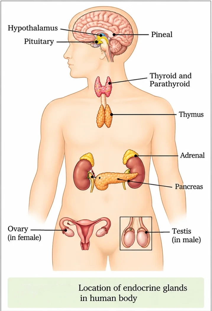
The hypothalamus
- The hypothalamus is the basal part of the diencephalon, forebrain, and it regulates a wide spectrum of body functions.
- It contains several groups of neurosecretory cells called nuclei, which produce hormones. These hormones regulate the synthesis and secretion of pituitary hormones.
- The hormones produced by the hypothalamus are of two types: the releasing hormones (which stimulate secretion of pituitary hormones) and the inhibiting hormones (which inhibit secretions of pituitary hormones). For example, gonadotrophin-releasing hormone (GnRH) stimulates the pituitary synthesis and release of gonadotrophins. On the other hand, somatostatin from the hypothalamus inhibits the release of growth hormone from the pituitary.
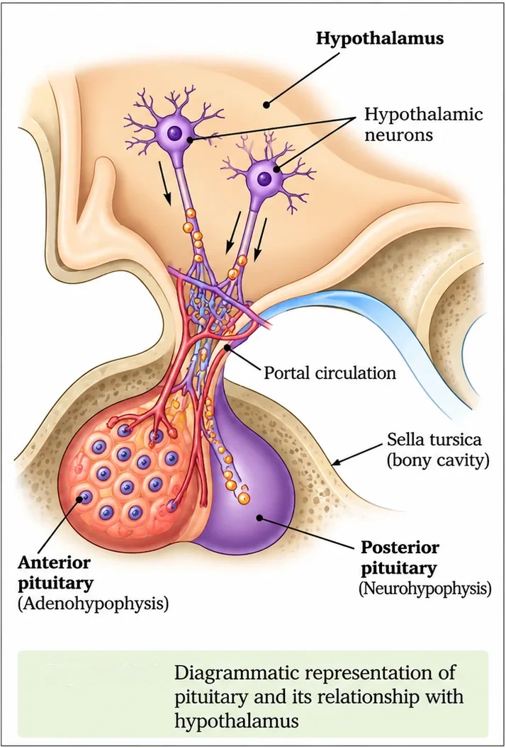
- These hormones originating in the hypothalamic neurons pass through axons and are released from their nerve endings. They reach the pituitary gland through a portal circulatory system and regulate the functions of the anterior pituitary. The posterior pituitary is under the direct neural regulation of the hypothalamus.
The pituitary gland
- The pituitary gland is located in a bony cavity called the sella turcica and is attached to the hypothalamus by a stalk.
- It is divided anatomically into an adenohypophysis and a neurohypophysis.
- The adenohypophysis consists of two portions, the pars distalis and the pars intermedia. The pars distalis region of the pituitary, commonly called the anterior pituitary, produces growth hormone (GH), prolactin (PRL), thyroid-stimulating hormone (TSH), adrenocorticotrophic hormone (ACTH), luteinizing hormone (LH) and follicle-stimulating hormone (FSH).
- The pars intermedia secretes only one hormone called melanocyte-stimulating hormone (MSH). However, in humans, the pars intermedia is almost merged with the pars distalis.
- The neurohypophysis (pars nervosa), also known as the posterior pituitary, stores and releases two hormones called oxytocin and vasopressin, which are actually synthesised by the hypothalamus and transported axonally to the neurohypophysis.
| Pituitary hormone | Main action | Disorder |
|---|---|---|
| Growth hormone (GH) | Growth of the body | Over-secretion: gigantism; low secretion: pituitary dwarfism. Excess in adults (especially middle age): acromegaly — severe disfigurement, especially of the face |
| Prolactin (PRL) | Growth of mammary glands and formation of milk | — |
| TSH | Stimulates synthesis and secretion of thyroid hormones from the thyroid gland | — |
| ACTH | Stimulates synthesis and secretion of steroid hormones called glucocorticoids from the adrenal cortex | — |
| LH and FSH (gonadotrophins) | In males, LH stimulates synthesis and secretion of androgens from testis; FSH and androgens regulate spermatogenesis. In females, LH induces ovulation of fully mature follicles (Graafian follicles) and maintains the corpus luteum; FSH stimulates growth and development of ovarian follicles | — |
| MSH | Acts on melanocytes (melanin-containing cells) and regulates pigmentation of the skin | — |
| Oxytocin | Acts on smooth muscles; stimulates a vigorous contraction of the uterus at the time of child birth, and milk ejection from the mammary gland | — |
| Vasopressin (ADH) | Acts mainly at the kidney and stimulates resorption of water and electrolytes by the distal tubules, thereby reducing loss of water through urine (diuresis) | Impairment: diabetes insipidus — loss of water and dehydration |
The pineal gland
The pineal gland is located on the dorsal side of the forebrain. It secretes a hormone called melatonin. Melatonin plays a very important role in the regulation of a 24-hour (diurnal) rhythm of our body. For example, it helps in maintaining the normal rhythms of the sleep–wake cycle and body temperature. In addition, melatonin also influences metabolism, pigmentation, the menstrual cycle and our defence capability.
The thyroid gland
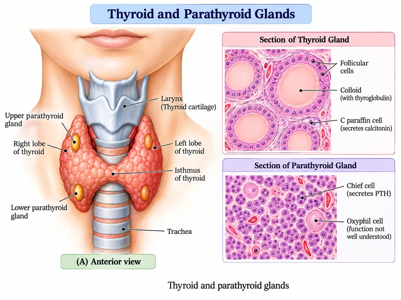
- The thyroid gland is composed of two lobes, located on either side of the trachea. Both lobes are interconnected with a thin flap of connective tissue called the isthmus.
- The thyroid gland is composed of follicles and stromal tissues. Each thyroid follicle is composed of follicular cells enclosing a cavity. These follicular cells synthesise two hormones, tetraiodothyronine or thyroxine (T₄) and triiodothyronine (T₃).
- Iodine is essential for the normal rate of hormone synthesis in the thyroid. Deficiency of iodine in our diet results in hypothyroidism and enlargement of the thyroid gland, commonly called goitre.
- Hypothyroidism during pregnancy causes defective development and maturation of the growing baby, leading to stunted growth (cretinism), mental retardation, low intelligence quotient, abnormal skin, deaf-mutism and so on. In adult women, hypothyroidism may cause the menstrual cycle to become irregular.
- Due to cancer of the thyroid gland or due to development of nodules of the thyroid glands, the rate of synthesis and secretion of thyroid hormones is increased to abnormally high levels, leading to a condition called hyperthyroidism, which adversely affects body physiology.
- Exophthalmic goitre (Graves' disease) is a form of hyperthyroidism characterised by enlargement of the thyroid gland, protrusion of the eyeballs, increased basal metabolic rate and weight loss.
- Thyroid hormones play an important role in the regulation of the basal metabolic rate. These hormones also support the process of red blood cell formation. Thyroid hormones control the metabolism of carbohydrates, proteins and fats. Maintenance of water and electrolyte balance is also influenced by thyroid hormones.
- The thyroid gland also secretes a protein hormone called thyrocalcitonin (TCT), which regulates the blood calcium levels.
The parathyroid gland
- In humans, four parathyroid glands are present on the back side of the thyroid gland, one pair each in the two lobes of the thyroid gland.
- The parathyroid glands secrete a peptide hormone called parathyroid hormone (PTH). The secretion of PTH is regulated by the circulating levels of calcium ions.
- PTH increases the Ca²⁺ levels in the blood. It acts on bones and stimulates the process of bone resorption (dissolution/demineralisation). PTH also stimulates reabsorption of Ca²⁺ by the renal tubules and increases Ca²⁺ absorption from the digested food.
- It is thus clear that PTH is a hypercalcemic hormone, i.e. it increases the blood Ca²⁺ levels. Along with TCT, it plays a significant role in calcium balance in the body.
The thymus
- The thymus gland is a lobular structure located on the dorsal side of the heart and the aorta.
- The thymus plays a major role in the development of the immune system. This gland secretes peptide hormones called thymosins.
- Thymosins play a major role in the differentiation of T-lymphocytes, which provide cell-mediated immunity. In addition, thymosins also promote production of antibodies to provide humoral immunity.
- The thymus is degenerated in old individuals, resulting in a decreased production of thymosins. As a result, the immune responses of old persons become weak.
The adrenal gland
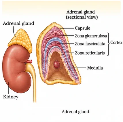
- Our body has one pair of adrenal glands, one at the anterior part of each kidney. The gland is composed of two types of tissues: the centrally located tissue called the adrenal medulla, and outside this lies the adrenal cortex.
- The adrenal medulla secretes two hormones called adrenaline or epinephrine and noradrenaline or norepinephrine. These are commonly called catecholamines.
- Adrenaline and noradrenaline are rapidly secreted in response to stress of any kind and during emergency situations, and are called emergency hormones or hormones of Fight or Flight.
- These hormones increase alertness, pupillary dilation, piloerection (raising of hairs), sweating and so on. Both the hormones increase the heart beat, the strength of heart contraction and the rate of respiration. Catecholamines also stimulate the breakdown of glycogen, resulting in an increased concentration of glucose in blood. In addition, they also stimulate the breakdown of lipids and proteins.
- The adrenal cortex can be divided into three layers, called the inner zona reticularis, the middle zona fasciculata and the outer zona glomerulosa.
- The adrenal cortex secretes many hormones, commonly called corticoids. The corticoids involved in carbohydrate metabolism are called glucocorticoids. In our body, cortisol is the main glucocorticoid. Corticoids which regulate the balance of water and electrolytes in our body are called mineralocorticoids. Aldosterone is the main mineralocorticoid in our body.
- Glucocorticoids stimulate gluconeogenesis, lipolysis and proteolysis, and inhibit cellular uptake and utilisation of amino acids. Cortisol is also involved in maintaining the cardiovascular system as well as the kidney functions. Glucocorticoids, particularly cortisol, produce anti-inflammatory reactions and suppress the immune response. Cortisol stimulates RBC production.
- Aldosterone acts mainly at the renal tubules and stimulates the reabsorption of Na⁺ and water and excretion of K⁺ and phosphate ions. Thus, aldosterone helps in the maintenance of electrolytes, body fluid volume, osmotic pressure and blood pressure.
- Small amounts of androgenic steroids are also secreted by the adrenal cortex, which play a role in the growth of axial hair, pubic hair and facial hair during puberty.
- Underproduction of hormones by the adrenal cortex alters carbohydrate metabolism, causing acute weakness and fatigue, leading to a disease called Addison's disease.
The pancreas
- The pancreas is a composite gland which acts as both exocrine and endocrine gland.
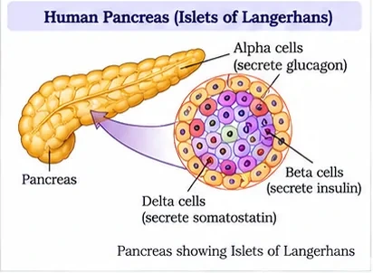
- The endocrine pancreas consists of Islets of Langerhans. There are about 1 to 2 million Islets of Langerhans in a normal human pancreas, representing only 1 to 2 per cent of the pancreatic tissue.
- The two main types of cells in the Islet of Langerhans are called α-cells and β-cells. The α-cells secrete a hormone called glucagon, while the β-cells secrete insulin.
- Glucagon is a peptide hormone and plays an important role in maintaining normal blood glucose levels. Glucagon acts mainly on the liver cells (hepatocytes) and stimulates glycogenolysis, resulting in an increased blood sugar (hyperglycemia). In addition, this hormone stimulates the process of gluconeogenesis, which also contributes to hyperglycemia. Glucagon reduces cellular glucose uptake and utilisation. Thus, glucagon is a hyperglycemic hormone.
- Insulin is a peptide hormone which plays a major role in the regulation of glucose homeostasis. Insulin acts mainly on hepatocytes and adipocytes (cells of adipose tissue) and enhances cellular glucose uptake and utilisation. As a result, there is a rapid movement of glucose from blood to hepatocytes and adipocytes, resulting in decreased blood glucose levels (hypoglycemia). Insulin also stimulates conversion of glucose to glycogen (glycogenesis) in the target cells.
- The glucose homeostasis in blood is thus maintained jointly by the two hormones — insulin and glucagon.
- Prolonged hyperglycemia leads to a complex disorder called diabetes mellitus, which is associated with loss of glucose through urine and formation of harmful compounds known as ketone bodies. Diabetic patients are successfully treated with insulin therapy.
Testis and ovary
- A pair of testes is present in the scrotal sac (outside the abdomen) of males. The testis performs dual functions as a primary sex organ as well as an endocrine gland.
- The testis is composed of seminiferous tubules and stromal or interstitial tissue. The Leydig cells or interstitial cells, which are present in the intertubular spaces, produce a group of hormones called androgens, mainly testosterone.
- Androgens regulate the development, maturation and functions of the male accessory sex organs like the epididymis, vas deferens, seminal vesicles, prostate gland, urethra and so on. These hormones stimulate muscular growth, growth of facial and axillary hair, aggressiveness and low pitch of voice. Androgens play a major stimulatory role in the process of spermatogenesis (formation of spermatozoa). Androgens act on the central neural system and influence male sexual behaviour (libido). These hormones produce anabolic (synthetic) effects on protein and carbohydrate metabolism.
- Females have a pair of ovaries located in the abdomen. The ovary is the primary female sex organ which produces one ovum during each menstrual cycle. In addition, the ovary also produces two groups of steroid hormones called estrogen and progesterone.
- The ovary is composed of ovarian follicles and stromal tissues. Estrogen is synthesised and secreted mainly by the growing ovarian follicles. After ovulation, the ruptured follicle is converted into a structure called the corpus luteum, which secretes mainly progesterone.
- Estrogens produce wide-ranging actions such as stimulation of growth and activities of female secondary sex organs, development of growing ovarian follicles, appearance of female secondary sex characters (e.g. high pitch of voice), mammary gland development. Estrogens also regulate female sexual behaviour.
- Progesterone supports pregnancy. Progesterone also acts on the mammary glands and stimulates the formation of alveoli (sac-like structures which store milk) and milk secretion.
4. Hormones of Heart, Kidney and Gastrointestinal Tract
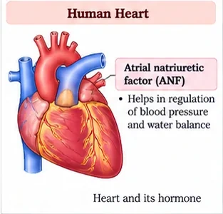
- The atrial wall of our heart secretes a very important peptide hormone called atrial natriuretic factor (ANF), which decreases blood pressure. When blood pressure is increased, ANF is secreted, which causes dilation of the blood vessels. This reduces the blood pressure.
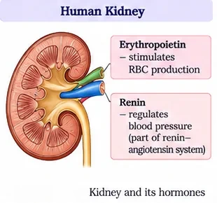
- The juxtaglomerular cells of the kidney produce a peptide hormone called erythropoietin, which stimulates erythropoiesis (formation of RBCs).
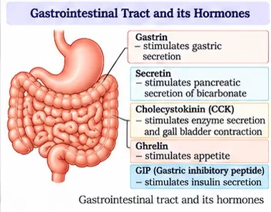
- Endocrine cells present in different parts of the gastrointestinal tract secrete four major peptide hormones, namely gastrin, secretin, cholecystokinin (CCK) and gastric inhibitory peptide (GIP).
- Gastrin acts on the gastric glands and stimulates the secretion of hydrochloric acid and pepsinogen. Secretin acts on the exocrine pancreas and stimulates secretion of water and bicarbonate ions. CCK acts on both pancreas and gall bladder and stimulates the secretion of pancreatic enzymes and bile juice, respectively. GIP inhibits gastric secretion and motility.
- Several other non-endocrine tissues secrete hormones called growth factors. These factors are essential for the normal growth of tissues and their repairing/regeneration.
5. Mechanism of Hormone Action
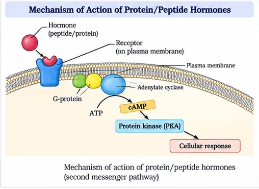
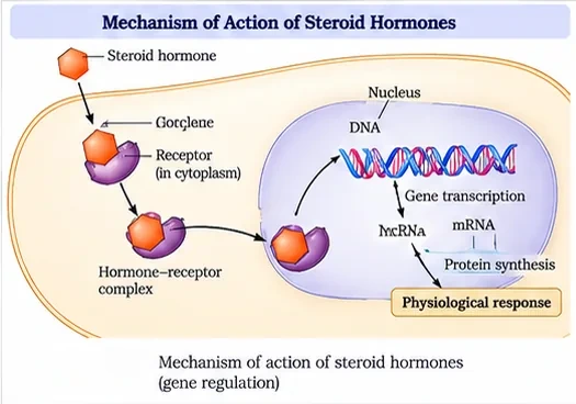
- Hormones produce their effects on target tissues by binding to specific proteins called hormone receptors located in the target tissues only.
- Hormone receptors present on the cell membrane of the target cells are called membrane-bound receptors, and the receptors present inside the target cell are called intracellular receptors, mostly nuclear receptors (present in the nucleus).
- Binding of a hormone to its receptor leads to the formation of a hormone–receptor complex. Each receptor is specific to one hormone only, and hence receptors are specific.
- Hormone–receptor complex formation leads to certain biochemical changes in the target tissue. Target tissue metabolism and hence physiological functions are regulated by hormones.
| Chemical nature | Examples | Receptor and action |
|---|---|---|
| Peptide, polypeptide, protein hormones | Insulin, glucagon, pituitary hormones, hypothalamic hormones | Do not enter the target cell; bind membrane-bound receptors and generate second messengers (e.g. cyclic AMP, IP₃, Ca²⁺), which in turn regulate cellular metabolism |
| Steroids | Cortisol, testosterone, estradiol, progesterone | Interact with intracellular receptors; mostly regulate gene expression or chromosome function by the interaction of the hormone–receptor complex with the genome |
| Iodothyronines | Thyroid hormones (T₃, T₄) | Intracellular receptors; regulate gene expression |
| Amino-acid derivatives | Epinephrine | Membrane receptors |
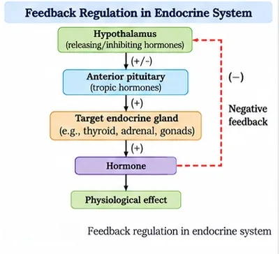
Most endocrine glands are regulated by feedback. For example, the hypothalamus releases a releasing hormone that stimulates the anterior pituitary to release a tropic hormone; this stimulates a target gland (e.g. thyroid), whose hormone, when it rises in blood, inhibits the hypothalamus and pituitary (negative feedback). This keeps hormone levels within a narrow range.
6. Master Summary Table
| Gland | Hormone(s) | Key action | Disorder |
|---|---|---|---|
| Hypothalamus | GnRH, somatostatin etc. | Controls pituitary | — |
| Anterior pituitary | GH, PRL, TSH, ACTH, LH, FSH | Growth, milk, thyroid, adrenal, gonads | Gigantism, dwarfism, acromegaly |
| Posterior pituitary | Oxytocin, vasopressin | Uterine contraction, milk ejection; water reabsorption | Diabetes insipidus |
| Pineal | Melatonin | Diurnal rhythm, sleep–wake cycle | — |
| Thyroid | T₃, T₄, thyrocalcitonin | BMR, RBC formation; lowers Ca²⁺ | Goitre, cretinism, Graves' disease |
| Parathyroid | PTH | Raises blood Ca²⁺ | — |
| Thymus | Thymosins | T-lymphocyte differentiation | Weak immunity in old age |
| Adrenal medulla | Adrenaline, noradrenaline | Fight or flight | — |
| Adrenal cortex | Cortisol, aldosterone, androgens | Glucose, Na⁺ and water balance | Addison's disease |
| Pancreas | Insulin (β), glucagon (α) | Lower / raise blood glucose | Diabetes mellitus |
| Testis / ovary | Testosterone / estrogen, progesterone | Sex characters, gametogenesis, pregnancy | — |
| Heart / kidney | ANF / erythropoietin | Lower BP / RBC formation | — |
| GI tract | Gastrin, secretin, CCK, GIP | Digestive secretions | — |
7. Mind Map
- Hypothalamus
- Pituitary (anterior, posterior)
- Pineal
- Thyroid
- Parathyroid
- Thymus
- Adrenal
- Pancreas
- Gonads
- ANF, erythropoietin, GI hormones
- Membrane receptors (cAMP)
- Intracellular receptors
8. Tips and Tricks
- Anterior pituitary hormones: "FLAT PiG" — FSH, LH, ACTH, TSH, Prolactin, GH.
- Posterior pituitary releases but does not make: "Oxytocin and Vasopressin — OVer from the hypothalamus".
- Adrenal cortex layers outside → in: "GFR" — glomerulosa (salt: aldosterone), fasciculata (sugar: cortisol), reticularis (sex: androgens). "Salt, Sugar, Sex".
- Calcium: PTH raises it ("Push up"), TCT lowers it ("Take down").
- Insulin lowers glucose ("In to cells"); glucagon raises it ("Glucose Gone from liver to blood").
- Saying oxytocin and vasopressin are synthesised in the posterior pituitary (they are made in the hypothalamus).
- Confusing diabetes insipidus (ADH) with diabetes mellitus (insulin).
- Calling PTH hypocalcemic (it is hypercalcemic).
- Thinking steroid hormones use cAMP as second messenger (they act on intracellular receptors).
- Mixing up gigantism (excess GH in children) and acromegaly (excess GH in adults).
9. Important One-liners
- Endocrine glands are ductless glands.
- The pituitary lies in the sella turcica.
- Somatostatin from the hypothalamus inhibits the release of growth hormone.
- In humans, the pars intermedia is almost merged with the pars distalis.
- Melatonin regulates the 24-hour (diurnal) rhythm.
- Iodine deficiency causes goitre.
- Graves' disease is a form of hyperthyroidism with protruding eyeballs.
- The thymus degenerates in old age, weakening immunity.
- Adrenaline and noradrenaline are called emergency hormones.
- Cortisol is the main glucocorticoid and aldosterone the main mineralocorticoid.
- Islets of Langerhans make up only 1–2% of pancreatic tissue.
- Leydig cells secrete testosterone; the corpus luteum secretes progesterone.
10. NEET-pattern Questions with Detailed Answers
These questions are original and based on concepts that NEET UG asks again and again.
Which hormone is called the hormone of "fight or flight"?
- Insulin
- Thyroxine
- Adrenaline
- Melatonin
Show solution
Adrenaline from the adrenal medulla is released in emergencies. It raises heart rate, alertness and blood glucose. The other three hormones are not emergency hormones.
Answer: (C)
Deficiency of iodine in the diet causes:
- Graves' disease
- Goitre
- Acromegaly
- Addison's disease
Show solution
Without enough iodine, the thyroid cannot make enough T₃ and T₄ (hypothyroidism), and it enlarges to form a goitre. Graves' disease is hyperthyroidism, acromegaly is caused by excess GH, and Addison's disease by underactivity of the adrenal cortex.
Answer: (B)
Oxytocin and vasopressin are:
- Synthesised and released by the anterior pituitary
- Synthesised by the hypothalamus and released from the posterior pituitary
- Synthesised by the posterior pituitary
- Secreted by the adrenal medulla
Show solution
Hypothalamic neurons make these hormones and carry them along their axons to the neurohypophysis, which stores and releases them. The posterior pituitary does not make them itself.
Answer: (B)
Excess secretion of growth hormone in adults, especially in middle age, causes:
- Gigantism
- Dwarfism
- Acromegaly
- Cretinism
Show solution
In adults, excess GH causes acromegaly, with disfigurement of the face. Gigantism results from excess GH in children, and dwarfism from too little GH. Cretinism is caused by hypothyroidism.
Answer: (C)
Which hormone increases the blood calcium level?
- Thyrocalcitonin
- Parathyroid hormone
- Insulin
- Glucagon
Show solution
PTH raises blood Ca²⁺ by causing bone resorption, increasing renal reabsorption and increasing absorption from food. Thyrocalcitonin does the opposite.
Answer: (B)
Which hormone acts on the exocrine pancreas and stimulates secretion of water and bicarbonate ions?
- Gastrin
- Secretin
- CCK
- GIP
Show solution
Secretin makes the pancreas release a watery, bicarbonate-rich juice. Gastrin acts on the gastric glands, CCK causes release of pancreatic enzymes and bile, and GIP inhibits gastric secretion.
Answer: (B)
Assertion (A): Steroid hormones mostly regulate gene expression.
Reason (R): Steroid hormones bind to intracellular receptors.
- Both A and R are true, and R explains A
- Both A and R are true, but R does not explain A
- A is true, R is false
- A is false, R is true
Show solution
Steroids are lipid-soluble, so they cross the cell membrane and bind receptors inside the cell. The hormone–receptor complex then acts on the genome. So R explains A.
Answer: (A)
Which statement about the adrenal cortex is correct?
- The zona reticularis is the outermost layer
- Aldosterone stimulates reabsorption of Na⁺ and excretion of K⁺
- Cortisol promotes the immune response
- Its underproduction causes Graves' disease
Show solution
Aldosterone acts on the renal tubules to retain Na⁺ and water and to excrete K⁺. The zona glomerulosa is the outermost layer, cortisol suppresses the immune response, and underproduction causes Addison's disease, not Graves' disease.
Answer: (B)
A hormone that does NOT enter its target cell and acts through cyclic AMP is:
- Cortisol
- Thyroxine
- Glucagon
- Progesterone
Show solution
Glucagon is a peptide hormone. It binds a membrane receptor and generates second messengers such as cAMP. Cortisol and progesterone (steroids) and thyroxine (an iodothyronine) act through intracellular receptors.
Answer: (C)
Match: (a) Melatonin (b) Thymosins (c) Erythropoietin (d) ANF — (i) Juxtaglomerular cells (ii) Atrial wall (iii) Pineal gland (iv) Thymus.
- a-iii, b-iv, c-i, d-ii
- a-iv, b-iii, c-ii, d-i
- a-iii, b-iv, c-ii, d-i
- a-i, b-iv, c-iii, d-ii
Show solution
Melatonin comes from the pineal gland, thymosins from the thymus, erythropoietin from the juxtaglomerular cells of the kidney, and ANF from the atrial wall of the heart.
Answer: (A)
11. Quick Revision Summary
- Hormone: non-nutrient intercellular messenger produced in trace amounts.
- Hypothalamus → releasing/inhibiting hormones → anterior pituitary (portal system); posterior pituitary stores oxytocin and vasopressin made in hypothalamus.
- Anterior pituitary: GH, PRL, TSH, ACTH, LH, FSH (+ MSH). GH disorders: gigantism, dwarfism, acromegaly. ADH deficiency: diabetes insipidus.
- Pineal: melatonin. Thyroid: T₃, T₄ (iodine; goitre, cretinism, Graves'), TCT. Parathyroid: PTH (raises Ca²⁺). Thymus: thymosins.
- Adrenal medulla: catecholamines (fight or flight). Cortex: cortisol, aldosterone, androgens; Addison's disease.
- Pancreas: β-cells insulin (lowers glucose), α-cells glucagon (raises); diabetes mellitus.
- Gonads: testosterone (Leydig cells); estrogen and progesterone (follicle, corpus luteum). Heart ANF, kidney erythropoietin, GI: gastrin, secretin, CCK, GIP.
- Peptide hormones → membrane receptors → second messengers (cAMP); steroids and iodothyronines → intracellular receptors → gene expression.