
1. Chapter at a Glance
The functions of the organs and organ systems in our body must be coordinated to maintain homeostasis. Coordination is the process through which two or more organs interact and complement each other's functions. For example, during physical exercise, the energy demand increases: the respiration rate, heart beat and blood flow all increase together. The neural system and the endocrine system jointly coordinate and integrate all the activities of the organs. The neural system provides an organised network of point-to-point connections for quick coordination, whereas the endocrine system provides chemical integration through hormones. This chapter covers the neural system of animals, the human neural system, the neuron, generation and conduction of the nerve impulse, transmission at synapses, the central nervous system (brain and its parts) and reflex action. NEET asks about two questions every year, usually on resting potential, the sodium–potassium pump, synapses and the functions of brain parts.
- Parts of a neuron; multipolar, bipolar and unipolar neurons; myelinated vs non-myelinated fibres.
- Resting membrane potential, Na⁺–K⁺ pump (3 Na⁺ out, 2 K⁺ in), depolarisation and action potential.
- Saltatory conduction; electrical vs chemical synapse; neurotransmitters.
- Forebrain (cerebrum, thalamus, hypothalamus), midbrain and hindbrain (pons, cerebellum, medulla) functions.
- Limbic system, corpus callosum, reflex arc.
2. Neural System
- The neural system of all animals is composed of highly specialised cells called neurons, which can detect, receive and transmit different kinds of stimuli.
- The neural organisation is very simple in lower invertebrates. For example, in Hydra it is composed of a network of neurons.
- The neural system is better organised in insects, where a brain is present along with a number of ganglia and neural tissues.
- Vertebrates have a more developed neural system.
Human neural system
- The human neural system is divided into two parts: the central neural system (CNS) and the peripheral neural system (PNS).
- The CNS includes the brain and the spinal cord and is the site of information processing and control.
- The PNS comprises all the nerves of the body associated with the CNS (brain and spinal cord).
- Nerve fibres of the PNS are of two types: afferent fibres, which transmit impulses from tissues/organs to the CNS, and efferent fibres, which transmit regulatory impulses from the CNS to the concerned peripheral tissues/organs.
- The PNS is divided into the somatic neural system (relays impulses from the CNS to skeletal muscles) and the autonomic neural system (transmits impulses from the CNS to the involuntary organs and smooth muscles of the body).
- The autonomic neural system is further classified into the sympathetic and parasympathetic neural systems.
- The visceral nervous system is the part of the PNS that comprises the whole complex of nerves, fibres, ganglia and plexuses by which impulses travel from the CNS to the viscera and from the viscera to the CNS.
3. Neuron: Structural and Functional Unit
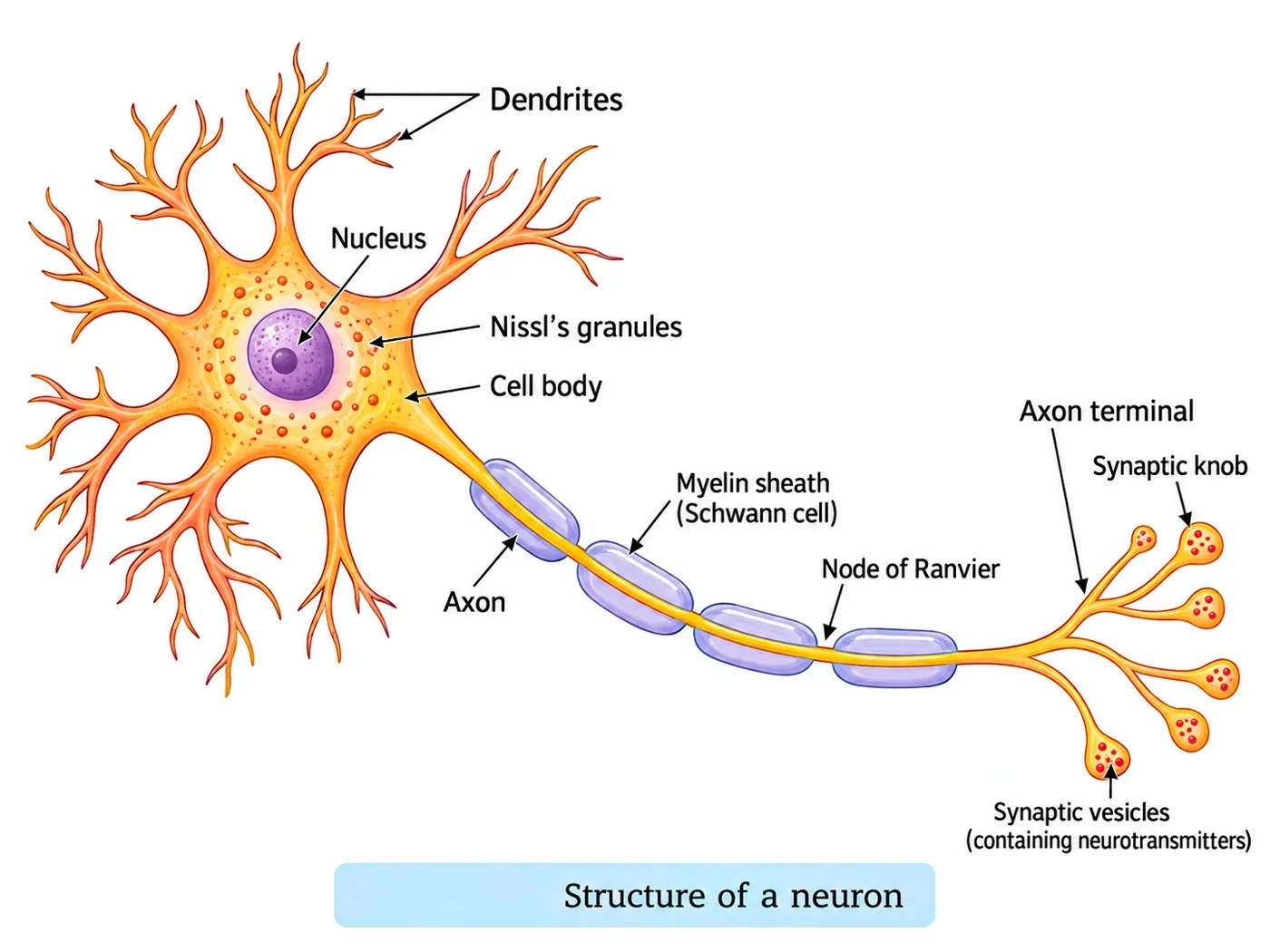
A neuron is a microscopic structure composed of three major parts — the cell body, dendrites and axon.
- The cell body contains cytoplasm with typical cell organelles and certain granular bodies called Nissl's granules.
- Short fibres which branch repeatedly and project out of the cell body also contain Nissl's granules and are called dendrites. These fibres transmit impulses towards the cell body.
- The axon is a long fibre, the distal end of which is branched. Each branch terminates as a bulb-like structure called the synaptic knob, which possesses synaptic vesicles containing chemicals called neurotransmitters. The axons transmit nerve impulses away from the cell body to a synapse or to a neuromuscular junction.
| Type (by number of axons and dendrites) | Structure | Found in |
|---|---|---|
| Multipolar | One axon and two or more dendrites | Cerebral cortex |
| Bipolar | One axon and one dendrite | Retina of the eye |
| Unipolar | Cell body with one axon only | Usually in the embryonic stage |
| Feature | Myelinated nerve fibres | Non-myelinated nerve fibres |
|---|---|---|
| Covering | Enveloped with Schwann cells, which form a myelin sheath around the axon | Enclosed by a Schwann cell that does not form a myelin sheath |
| Nodes of Ranvier | Gaps between two adjacent myelin sheaths | Absent |
| Found in | Spinal and cranial nerves | Autonomous and somatic neural systems |
| NEET Note | Saltatory conduction (faster) | Slower conduction |
4. Generation and Conduction of Nerve Impulse
Neurons are excitable cells because their membranes are in a polarised state. Different types of ion channels are present on the neural membrane; these are selectively permeable to different ions.
Resting potential
- When a neuron is not conducting any impulse (resting), the axonal membrane is comparatively more permeable to potassium ions (K⁺) and nearly impermeable to sodium ions (Na⁺). Similarly, it is impermeable to the negatively charged proteins present in the axoplasm.
- Consequently, the axoplasm inside the axon contains a high concentration of K⁺ and negatively charged proteins and a low concentration of Na⁺. In contrast, the fluid outside the axon contains a low concentration of K⁺, a high concentration of Na⁺, and thus forms a concentration gradient.
- These ionic gradients across the resting membrane are maintained by the active transport of ions by the sodium–potassium pump, which transports 3 Na⁺ outwards for 2 K⁺ into the cell.
- As a result, the outer surface of the axonal membrane possesses a positive charge while its inner surface becomes negatively charged, and therefore it is polarised. The electrical potential difference across the resting plasma membrane is called the resting potential (about −70 mV).
Action potential and conduction
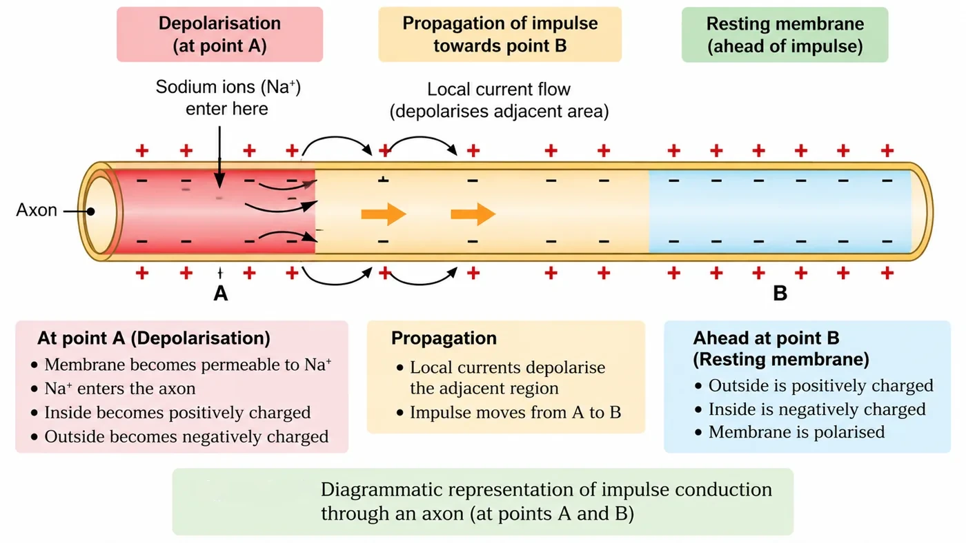
- When a stimulus is applied at a site (e.g. point A) on the polarised membrane, the membrane at site A becomes freely permeable to Na⁺. This leads to a rapid influx of Na⁺, followed by the reversal of the polarity at that site — the outer surface of the membrane becomes negatively charged and the inner side positively charged. The polarity of the membrane at site A is thus reversed and is said to be depolarised.
- The electrical potential difference across the plasma membrane at site A is called the action potential, which is in fact termed a nerve impulse.
- At sites immediately ahead (e.g. site B), the axon membrane has a positive charge on the outer surface and a negative charge on its inner surface. As a result, a current flows on the inner surface from site A to site B. On the outer surface, current flows from site B to site A to complete the circuit of current flow. Hence the polarity at site B is reversed, and an action potential is generated there. Thus the impulse (action potential) generated at site A arrives at site B. The sequence is repeated along the length of the axon, and consequently the impulse is conducted.
- The rise in the stimulus-induced permeability to Na⁺ is extremely short-lived. It is quickly followed by a rise in permeability to K⁺. Within a fraction of a second, K⁺ diffuses outside the membrane and restores the resting potential of the membrane at the site of excitation, and the fibre becomes once more responsive to further stimulation (repolarisation).
5. Transmission of Impulses
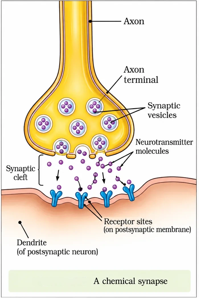
A nerve impulse is transmitted from one neuron to another through junctions called synapses. A synapse is formed by the membranes of a pre-synaptic neuron and a post-synaptic neuron, which may or may not be separated by a gap called the synaptic cleft. There are two types of synapses — electrical and chemical.
| Feature | Electrical synapse | Chemical synapse |
|---|---|---|
| Membranes | Pre- and post-synaptic membranes are in very close proximity | Separated by a fluid-filled space, the synaptic cleft |
| How impulse crosses | Electric current flows directly from one neuron into the other | Neurotransmitters carry the signal across the cleft |
| Speed | Always faster | Slower |
| Occurrence | Rare in our system | Common |
- At a chemical synapse, when an impulse (action potential) arrives at the axon terminal, it stimulates the movement of the synaptic vesicles towards the membrane, where they fuse with the plasma membrane and release their neurotransmitters into the synaptic cleft.
- The released neurotransmitters bind to their specific receptors present on the post-synaptic membrane. This binding opens ion channels, allowing the entry of ions that can generate a new potential in the post-synaptic neuron.
- The new potential developed may be either excitatory or inhibitory.
- Acetylcholine is an example of a neurotransmitter.
6. Central Neural System
The brain is the central information-processing organ of our body and acts as the "command and control system". It controls voluntary movements, balance of the body, functioning of vital involuntary organs (e.g. lungs, heart, kidneys), thermoregulation, hunger and thirst, circadian (24-hour) rhythms of our body, activities of several endocrine glands and human behaviour. It is also the site for processing of vision, hearing, speech, memory, intelligence, emotions and thoughts.
- The human brain is well protected by the skull. Inside the skull, the brain is covered by cranial meninges consisting of an outer layer called the dura mater, a very thin middle layer called the arachnoid, and an inner layer (which is in contact with the brain tissue) called the pia mater.
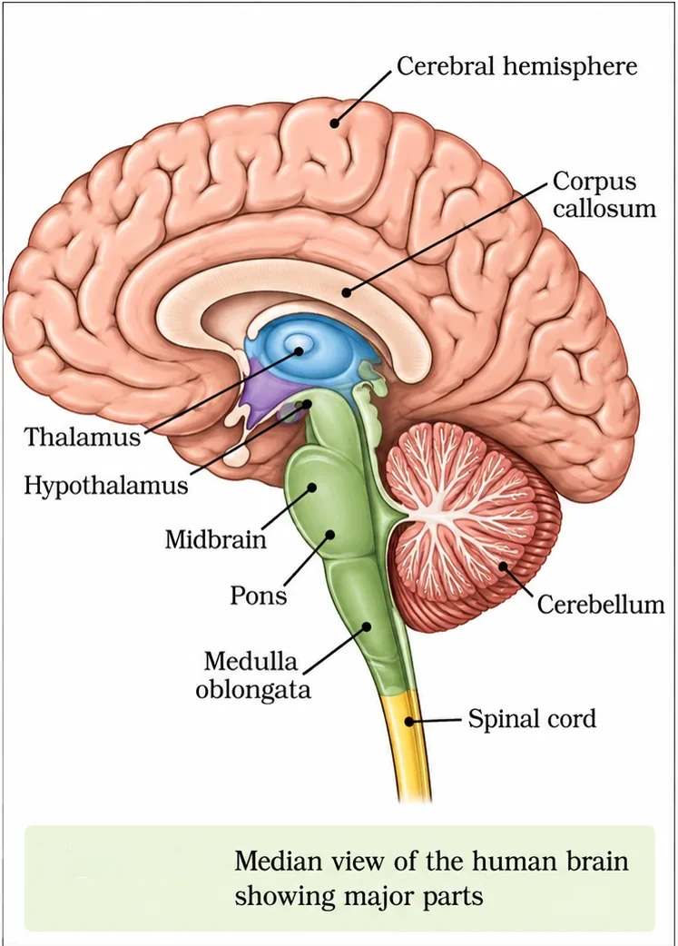
- The brain can be divided into three major parts: (i) forebrain, (ii) midbrain and (iii) hindbrain.
Forebrain
- The forebrain consists of the cerebrum, thalamus and hypothalamus.
- The cerebrum forms the major part of the human brain. A deep cleft divides it longitudinally into two halves, termed the left and right cerebral hemispheres. The hemispheres are connected by a tract of nerve fibres called the corpus callosum.
- The layer of cells which covers the cerebral hemisphere is called the cerebral cortex; it is thrown into prominent folds. The cerebral cortex is referred to as the grey matter due to its greyish appearance; the neuron cell bodies are concentrated here, giving the colour.
- The cerebral cortex contains motor areas, sensory areas and large regions that are neither clearly sensory nor motor in function. These regions, called the association areas, are responsible for complex functions like intersensory associations, memory and communication.
- Fibres of the tracts are covered with the myelin sheath, which constitute the inner part of the cerebral hemisphere. They give an opaque white appearance to the layer and, hence, it is called the white matter.
- The cerebrum wraps around a structure called the thalamus, which is a major coordinating centre for sensory and motor signalling.
- Another very important part of the brain called the hypothalamus lies at the base of the thalamus. The hypothalamus contains a number of centres which control body temperature, the urge for eating and drinking. It also contains several groups of neurosecretory cells which secrete hormones called hypothalamic hormones.
- The inner parts of the cerebral hemispheres and a group of associated deep structures like the amygdala, hippocampus and so on form a complex structure called the limbic lobe or limbic system. Along with the hypothalamus, it is involved in the regulation of sexual behaviour, expression of emotional reactions (e.g. excitement, pleasure, rage and fear) and motivation.
Midbrain
- The midbrain is located between the thalamus/hypothalamus of the forebrain and the pons of the hindbrain.
- A canal called the cerebral aqueduct passes through the midbrain.
- The dorsal portion of the midbrain consists mainly of four round swellings (lobes) called corpora quadrigemina.
- The midbrain and hindbrain form the brain stem.
Hindbrain
- The hindbrain comprises the pons, cerebellum and medulla (also called the medulla oblongata).
- The pons consists of fibre tracts that interconnect different regions of the brain.
- The cerebellum has a very convoluted surface in order to provide the additional space for many more neurons.
- The medulla of the brain is connected to the spinal cord. The medulla contains centres which control respiration, cardiovascular reflexes and gastric secretions.
- Three major regions make up the brain stem: midbrain, pons and medulla oblongata. The brain stem forms the connection between the brain and the spinal cord.
| Part of brain | Region | Main function |
|---|---|---|
| Cerebrum | Forebrain | Thinking, memory, intelligence, voluntary actions, sensory and motor areas |
| Corpus callosum | Forebrain | Connects the two cerebral hemispheres |
| Thalamus | Forebrain | Major coordinating centre for sensory and motor signalling |
| Hypothalamus | Forebrain | Body temperature, hunger, thirst; secretes hypothalamic hormones |
| Limbic system | Forebrain | Emotions, sexual behaviour, motivation |
| Corpora quadrigemina | Midbrain | Visual and auditory reflexes |
| Pons | Hindbrain | Fibre tracts connecting brain regions; pneumotaxic centre |
| Cerebellum | Hindbrain | Balance, posture and coordination of movements |
| Medulla oblongata | Hindbrain | Respiration, cardiovascular reflexes, gastric secretion |
7. Reflex Action and Reflex Arc
- A reflex action is an entire process of response to a peripheral nervous stimulation that occurs involuntarily, i.e. without conscious effort or thought, and requires the involvement of a part of the central nervous system. The best-known example is the knee-jerk reflex.
- The reflex pathway comprises at least one afferent neuron (receptor) and one efferent (effector or excitor) neuron appropriately arranged in a series.
- The afferent neuron receives a signal from a sensory organ and transmits the impulse via a dorsal nerve root into the CNS (at the level of the spinal cord).
- The efferent neuron then carries signals from the CNS to the effector. The stimulus and response thus form a reflex arc.
8. Master Summary Table
| Topic | Key fact | NEET Note |
|---|---|---|
| Hydra | Network of neurons | Simplest neural organisation |
| Dendrites / axon | Towards / away from cell body | Nissl's granules in cell body and dendrites |
| Bipolar neurons | One axon, one dendrite | Retina |
| Resting potential | Outside +, inside − | Inside high K⁺, outside high Na⁺ |
| Na⁺–K⁺ pump | 3 Na⁺ out, 2 K⁺ in | Active transport |
| Depolarisation | Na⁺ influx | Polarity reversed |
| Repolarisation | K⁺ efflux | Resting state restored |
| Electrical synapse | Faster, rare | Chemical synapse uses neurotransmitters |
| Meninges | Dura, arachnoid, pia (outer to inner) | Pia touches brain |
| Hypothalamus | Temperature, hunger, thirst | Neurosecretory cells |
| Medulla | Respiration, heart, gastric secretion | Vital centres |
| Reflex arc | Afferent → CNS → efferent | Knee jerk |
9. Mind Map
- CNS, PNS
- Somatic, autonomic
- Afferent, efferent
- Structure, types
- Resting potential
- Action potential
- Electrical
- Chemical
- Neurotransmitters
- Fore-, mid-, hindbrain
- Limbic system
- Reflex arc
10. Tips and Tricks
- Sodium–potassium pump: "3 Salty Out, 2 Potatoes In" — 3 Na⁺ out, 2 K⁺ in.
- Meninges from outside: "Don't Act Proud" — dura, arachnoid, pia.
- "Na in = depolarise; K out = repolarise."
- Hindbrain "PCM" — pons, cerebellum, medulla.
- Dendrites "D for Deliver to cell body"; axon "A for Away".
- Saying the resting membrane is more permeable to Na⁺ (it is more permeable to K⁺).
- Reversing the pump ratio (it is 3 Na⁺ out for 2 K⁺ in).
- Calling electrical synapses common in humans (they are rare).
- Placing the respiratory centre in the cerebellum (it is in the medulla).
- Saying grey matter is the inner layer of the cerebrum (it is the outer cortex).
11. Important One-liners
- The neural system of Hydra is a network of neurons.
- Nissl's granules are found in the cell body and dendrites of a neuron.
- Bipolar neurons are found in the retina.
- Gaps between adjacent myelin sheaths are called nodes of Ranvier.
- The resting membrane is polarised: positive outside, negative inside.
- Electrical synapses conduct impulses faster than chemical synapses.
- The corpus callosum connects the two cerebral hemispheres.
- The hypothalamus controls body temperature, hunger and thirst.
- Corpora quadrigemina are four lobes on the dorsal side of the midbrain.
- The medulla oblongata contains centres controlling respiration and heart reflexes.
- The knee-jerk is an example of a reflex action.
12. NEET-pattern Questions with Detailed Answers
These questions are original and based on concepts that NEET UG asks again and again.
The sodium–potassium pump transports:
- 2 Na⁺ out and 3 K⁺ in
- 3 Na⁺ out and 2 K⁺ in
- 3 Na⁺ in and 2 K⁺ out
- Equal numbers of Na⁺ and K⁺
Show solution
The pump uses ATP to move 3 Na⁺ out for every 2 K⁺ it brings in. This keeps the inside negative. The other options reverse the ratio or the direction.
Answer: (B)
Bipolar neurons are found in the:
- Cerebral cortex
- Retina
- Spinal cord
- Embryo only
Show solution
Bipolar neurons (one axon, one dendrite) occur in the retina. Multipolar neurons occur in the cerebral cortex, and unipolar neurons are usually found in the embryo.
Answer: (B)
In a resting neuron, the axonal membrane is:
- More permeable to Na⁺ than K⁺
- More permeable to K⁺ and nearly impermeable to Na⁺
- Equally permeable to Na⁺ and K⁺
- Impermeable to both
Show solution
At rest, K⁺ channels allow some K⁺ to move, while Na⁺ is largely kept out, which keeps the membrane polarised. Na⁺ permeability rises only when the membrane is stimulated.
Answer: (B)
The centres controlling respiration, cardiovascular reflexes and gastric secretion are located in the:
- Cerebellum
- Hypothalamus
- Medulla oblongata
- Corpus callosum
Show solution
The medulla, which connects to the spinal cord, holds these vital centres. The cerebellum coordinates movement, the hypothalamus controls temperature, hunger and thirst, and the corpus callosum joins the two hemispheres.
Answer: (C)
The structure that connects the two cerebral hemispheres is the:
- Pons
- Corpus callosum
- Corpora quadrigemina
- Thalamus
Show solution
The corpus callosum is a tract of nerve fibres joining the left and right hemispheres. The pons connects other brain regions, the corpora quadrigemina lie in the midbrain, and the thalamus is a sensory and motor relay centre.
Answer: (B)
When a nerve fibre is stimulated, depolarisation is caused by:
- Efflux of K⁺
- Influx of Na⁺
- Influx of Cl⁻
- Efflux of Na⁺
Show solution
The stimulus makes the membrane freely permeable to Na⁺. Na⁺ rushes in and reverses the polarity. K⁺ efflux comes afterwards and causes repolarisation.
Answer: (B)
Assertion (A): Impulse transmission across an electrical synapse is faster than across a chemical synapse.
Reason (R): In an electrical synapse, current flows directly from one neuron to the next.
- Both A and R are true, and R explains A
- Both A and R are true, but R does not explain A
- A is true, R is false
- A is false, R is true
Show solution
The membranes of an electrical synapse are very close together, and current passes directly without any neurotransmitter step. This is why it is faster, so R explains A.
Answer: (A)
Which structure, along with the hypothalamus, regulates emotions such as rage and fear and sexual behaviour?
- Cerebellum
- Limbic system
- Medulla
- Corpora quadrigemina
Show solution
The limbic system (including the amygdala and hippocampus) works with the hypothalamus to control emotions, sexual behaviour and motivation. The other three structures have different roles.
Answer: (B)
The correct sequence of the cranial meninges from outside to inside is:
- Pia mater, arachnoid, dura mater
- Dura mater, arachnoid, pia mater
- Arachnoid, dura mater, pia mater
- Dura mater, pia mater, arachnoid
Show solution
The tough dura mater is outermost, the thin arachnoid is in the middle, and the pia mater lies directly on the brain tissue. Option (A) is the reverse order.
Answer: (B)
Match: (a) Thalamus (b) Cerebellum (c) Pons (d) Hypothalamus — (i) Fibre tracts interconnecting brain regions (ii) Major coordinating centre for sensory and motor signals (iii) Highly convoluted surface for more neurons (iv) Thermoregulation and neurosecretion.
- a-ii, b-iii, c-i, d-iv
- a-iii, b-ii, c-iv, d-i
- a-ii, b-i, c-iii, d-iv
- a-iv, b-iii, c-i, d-ii
Show solution
The thalamus coordinates sensory and motor signals. The cerebellum has a convoluted surface to fit more neurons. The pons carries fibre tracts. The hypothalamus controls temperature and has neurosecretory cells.
Answer: (A)
13. Quick Revision Summary
- Neural and endocrine systems coordinate the body; Hydra has a nerve net.
- CNS (brain, spinal cord) + PNS (somatic and autonomic: sympathetic, parasympathetic); afferent vs efferent fibres.
- Neuron: cell body (Nissl's granules), dendrites (towards), axon (away); multipolar, bipolar (retina), unipolar.
- Resting: outside +, inside −; Na⁺–K⁺ pump 3 out : 2 in. Stimulus → Na⁺ influx (depolarisation) → K⁺ efflux (repolarisation).
- Synapse: electrical (fast, rare) vs chemical (neurotransmitters such as acetylcholine).
- Brain: meninges (dura, arachnoid, pia); forebrain (cerebrum, corpus callosum, thalamus, hypothalamus, limbic system), midbrain (corpora quadrigemina), hindbrain (pons, cerebellum, medulla).
- Reflex arc: receptor → afferent → spinal cord → efferent → effector (knee jerk).