
1. Chapter at a Glance
Movement is one of the significant features of living beings. Animals and plants show a wide range of movements: streaming of protoplasm in Amoeba, movement of cilia and flagella, and movement of limbs, jaws, eyelids and tongue. Some movements result in a change of place or location; such voluntary movements are called locomotion (walking, running, climbing, flying, swimming). All locomotions are movements, but all movements are not locomotions. This chapter covers the types of movement, the structure of skeletal muscle and the sliding filament mechanism of contraction, the human skeletal system, joints, and disorders of the muscular and skeletal systems. NEET asks about two questions every year, usually on the sarcomere, muscle proteins, the role of Ca²⁺, the number of bones and types of joints.
- Amoeboid, ciliary and muscular movement with examples.
- Skeletal, visceral and cardiac muscles; structure of a muscle fibre and sarcomere.
- Actin (with troponin and tropomyosin) and myosin (heavy and light meromyosin).
- Steps of contraction: neuromuscular junction, acetylcholine, Ca²⁺, cross bridge, ATP, sliding filament theory.
- 206 bones: axial and appendicular skeleton, number of vertebrae, true/false/floating ribs.
- Fibrous, cartilaginous and synovial joints; myasthenia gravis, tetany, muscular dystrophy, arthritis, osteoporosis, gout.
2. Types of Movement
Cells of the human body exhibit three main types of movements — amoeboid, ciliary and muscular.
| Type | How it happens | Examples in humans |
|---|---|---|
| Amoeboid | Pseudopodia formed by the streaming of protoplasm (as in Amoeba); cytoskeletal elements like microfilaments are also involved | Macrophages and leucocytes in blood |
| Ciliary | Coordinated movement of cilia | Removal of dust particles and foreign substances inhaled along with air from the trachea; passage of ova through the female reproductive tract |
| Muscular | Contractile property of muscles | Movement of limbs, jaws, tongue |
Locomotion requires a perfect coordinated activity of the muscular, skeletal and neural systems.
3. Muscle
Muscle is a specialised tissue of mesodermal origin. About 40–50% of the body weight of a human adult is contributed by muscles. They have special properties like excitability, contractility, extensibility and elasticity.
| Feature | Skeletal | Visceral | Cardiac |
|---|---|---|---|
| Location | Closely associated with skeletal components | Inner walls of hollow visceral organs (alimentary canal, reproductive tract) | Heart |
| Appearance | Striated (striped) | Non-striated (smooth) | Striated |
| Control | Voluntary | Involuntary | Involuntary |
| Function | Locomotion and changes of body posture | Transport of food through the digestive tract and gametes through the genital tract | Pumping blood; cells assemble in a branching pattern |
Structure of a skeletal muscle
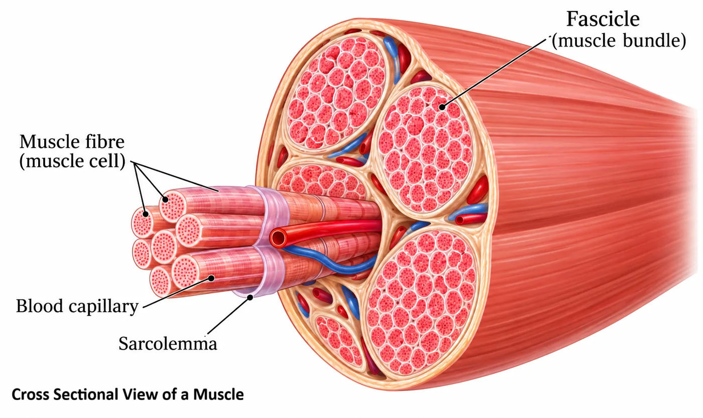
- Each organised skeletal muscle in our body is made of a number of muscle bundles or fascicles held together by a common collagenous connective tissue layer called fascia.
- Each muscle bundle contains a number of muscle fibres. Each muscle fibre is lined by the plasma membrane called sarcolemma, enclosing the sarcoplasm.
- A muscle fibre is a syncytium, as the sarcoplasm contains many nuclei.
- The endoplasmic reticulum of the muscle fibre is the sarcoplasmic reticulum, the store house of calcium ions.
- A characteristic feature of the muscle fibre is the presence of a large number of parallelly arranged filaments in the sarcoplasm called myofilaments or myofibrils.
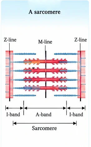
- Each myofibril has alternate dark and light bands on it. A detailed study has shown that the striated appearance is due to the distribution pattern of two important proteins — actin and myosin.
The sarcomere
- The light bands contain actin and are called I-bands or isotropic bands, whereas the dark bands contain myosin and are called A-bands or anisotropic bands.
- Both proteins are arranged as rod-like structures, parallel to each other and to the longitudinal axis of the myofibrils. Actin filaments are thinner than the myosin filaments, hence are commonly called thin and thick filaments respectively.
- In the centre of each I-band is an elastic fibre called the Z-line, which bisects it. The thin filaments are firmly attached to the Z-line.
- The thick filaments in the A-band are held together in the middle of this band by a thin fibrous membrane called the M-line.
- The A and I bands are arranged alternately throughout the length of the myofibrils.
- The portion of the myofibril between two successive Z-lines is considered the functional unit of contraction and is called a sarcomere.
- In a resting state, the edges of thin filaments on either side of the thick filaments partially overlap the free ends of the thick filaments, leaving the central part of the thick filaments. This central part of the thick filament, not overlapped by thin filaments, is called the H-zone.
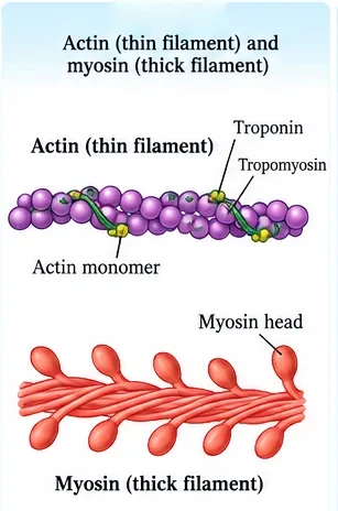
Structure of contractile proteins
- Each actin (thin) filament is made of two F (filamentous) actins helically wound to each other. Each F-actin is a polymer of monomeric G (globular) actins.
- Two filaments of another protein, tropomyosin, also run close to the F-actins throughout their length.
- A complex protein, troponin, is distributed at regular intervals on the tropomyosin. In the resting state, a subunit of troponin masks the active binding sites for myosin on the actin filaments.
- Each myosin (thick) filament is also a polymerised protein. Many monomeric proteins called meromyosins constitute one thick filament.
- Each meromyosin has two important parts: a globular head with a short arm and a tail. The former is called the heavy meromyosin (HMM) and the latter the light meromyosin (LMM).
- The HMM component, i.e. the head and short arm, projects outwards at regular distance and angle from each other from the surface of a polymerised myosin filament and is known as the cross arm.
- The globular head is an active ATPase enzyme and has binding sites for ATP and active sites for actin.
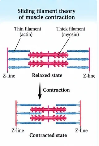
4. Mechanism of Muscle Contraction
The mechanism of muscle contraction is best explained by the sliding filament theory, which states that contraction of a muscle fibre takes place by the sliding of the thin filaments over the thick filaments.
- Muscle contraction is initiated by a signal sent by the CNS via a motor neuron. A motor neuron along with the muscle fibres connected to it constitutes a motor unit.
- The junction between a motor neuron and the sarcolemma of the muscle fibre is called the neuromuscular junction or motor-end plate.
- A neural signal reaching this junction releases a neurotransmitter (acetylcholine), which generates an action potential in the sarcolemma.
- This spreads through the muscle fibre and causes the release of calcium ions into the sarcoplasm.
- Increase in Ca²⁺ level leads to the binding of calcium with a subunit of troponin on actin filaments, thereby removing the masking of active sites for myosin.
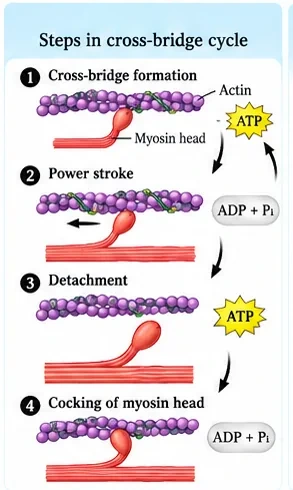
- Utilising the energy from ATP hydrolysis, the myosin head now binds to the exposed active sites on actin to form a cross bridge.
- This pulls the attached actin filaments towards the centre of the A-band. The Z-lines attached to these actins are also pulled inwards, thereby causing a shortening of the sarcomere, i.e. contraction.
- During shortening of the muscle (contraction), the I-bands get reduced, whereas the A-bands retain the length.
- The myosin, releasing the ADP and Pi, goes back to its relaxed state. A new ATP binds and the cross bridge is broken. The ATP is again hydrolysed by the myosin head, and the cycle of cross-bridge formation and breakage is repeated, causing further sliding.
- The process continues till the Ca²⁺ ions are pumped back to the sarcoplasmic cisternae, resulting in the masking of actin filaments. This causes the Z-lines to return to their original position, i.e. relaxation.
- The reaction time of the fibres can vary in different muscles. Repeated activation of the muscles can lead to the accumulation of lactic acid due to anaerobic breakdown of glycogen in them, causing fatigue.
- Muscle contains a red-coloured oxygen-storing pigment called myoglobin. Myoglobin content is high in some muscles, which gives a reddish appearance; such muscles are called red fibres. They contain plenty of mitochondria, which can utilise the large amount of oxygen stored in them for ATP production. These muscles can therefore also be called aerobic muscles.
- Some muscles possess a very small quantity of myoglobin and therefore appear pale or whitish. These are white fibres. The number of mitochondria is also few in them, but the amount of sarcoplasmic reticulum is high. They depend on anaerobic processes for energy.
| Part of sarcomere | During contraction | NEET Note |
|---|---|---|
| A-band | Length remains the same | Length of thick filament |
| I-band | Shortens | Thin filaments slide inwards |
| H-zone | Shortens or disappears | — |
| Sarcomere (Z to Z) | Shortens | Z-lines pulled closer |
| Thick and thin filaments | Lengths unchanged | They only slide |
5. Skeletal System
The skeletal system consists of a framework of bones and a few cartilages. This system has a significant role in movement shown by the body. Bone and cartilage are specialised connective tissues. The former has a very hard matrix due to calcium salts in it, and the latter has a slightly pliable matrix due to chondroitin salts. In human beings, this system is made up of 206 bones and a few cartilages. It is grouped into two principal divisions — the axial and the appendicular skeleton.
Axial skeleton (80 bones)
- Bones distributed along the main axis of the body: the skull, vertebral column, sternum and ribs.
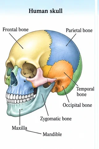
- The skull is composed of two sets of bones — cranial and facial — totalling 22 bones. Cranial bones are 8 in number; they form the hard protective outer covering, the cranium, for the brain. The facial region is made up of 14 skeletal elements which form the front part of the skull.
- A single U-shaped bone called the hyoid is present at the base of the buccal cavity, and it is also included in the skull.
- Each middle ear contains three tiny bones — malleus, incus and stapes — collectively called ear ossicles.
- The skull region articulates with the superior region of the vertebral column with the help of two occipital condyles (dicondylic skull).
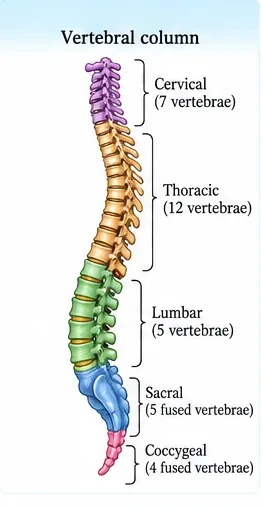
- Our vertebral column is formed by 26 serially arranged units called vertebrae and is dorsally placed. It extends from the base of the skull and constitutes the main framework of the trunk.
- Each vertebra has a central hollow portion (neural canal) through which the spinal cord passes. The first vertebra is the atlas, and it articulates with the occipital condyles.
- The vertebral column is differentiated into cervical (7), thoracic (12), lumbar (5), sacral (1, fused) and coccygeal (1, fused) regions starting from the skull. The number of cervical vertebrae is seven in almost all mammals, including humans.
- The vertebral column protects the spinal cord, supports the head and serves as the point of attachment for the ribs and musculature of the back.
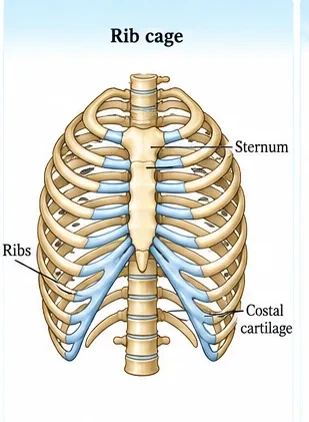
- The sternum is a flat bone on the ventral midline of the thorax.
- There are 12 pairs of ribs. Each rib is a thin flat bone connected dorsally to the vertebral column and ventrally to the sternum. It has two articulation surfaces on its dorsal end and is hence called bicephalic.
- The first seven pairs of ribs are called true ribs. Dorsally, they are attached to the thoracic vertebrae, and ventrally connected to the sternum with the help of hyaline cartilage.
- The 8th, 9th and 10th pairs of ribs do not articulate directly with the sternum but join the seventh rib with the help of hyaline cartilage. These are called vertebrochondral (false) ribs.
- The last two pairs (11th and 12th) of ribs are not connected ventrally and are therefore called floating ribs.
- The thoracic vertebrae, ribs and sternum together form the rib cage.
Appendicular skeleton (126 bones)
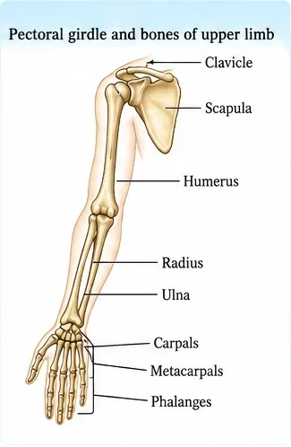
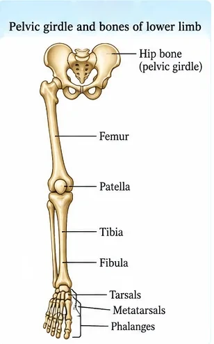
- The bones of the limbs along with their girdles constitute the appendicular skeleton. Each limb is made of 30 bones.
- Hand (fore limb): humerus, radius and ulna, carpals (wrist bones — 8 in number), metacarpals (palm bones — 5 in number) and phalanges (digits — 14 in number).
- Leg (hind limb): femur (thigh bone — the longest bone), tibia and fibula, tarsals (ankle bones — 7 in number), metatarsals (5 in number) and phalanges (digits — 14 in number). A cup-shaped bone called the patella covers the knee ventrally (knee cap).
- Pectoral and pelvic girdle bones help in the articulation of the upper and lower limbs respectively with the axial skeleton. Each girdle is formed of two halves.
- Each half of the pectoral girdle consists of a clavicle and a scapula. The scapula is a large triangular flat bone situated in the dorsal part of the thorax between the second and seventh ribs. Its expanded end is the acromion, which articulates with the clavicle. Below the acromion is a depression called the glenoid cavity, which articulates with the head of the humerus to form the shoulder joint. Each clavicle is a long slender bone with two curvatures, commonly called the collar bone.
- The pelvic girdle consists of two coxal bones. Each coxal bone is formed by the fusion of three bones — ilium, ischium and pubis. At the point of fusion of the above bones is a cavity called the acetabulum, to which the thigh bone articulates. The two halves of the pelvic girdle meet ventrally to form the pubic symphysis, containing fibrous cartilage.
6. Joints
Joints are essential for all types of movements involving the bony parts of the body. Locomotory movements are no exception. Joints are points of contact between bones, or between bones and cartilages. Force generated by the muscles is used to carry out movement through joints, where the joint acts as a fulcrum.
| Type | Description | Examples |
|---|---|---|
| Fibrous joints | Do not allow any movement. Bones are fused end to end with the help of dense fibrous connective tissues in the form of sutures. | Flat skull bones fusing to form the cranium |
| Cartilaginous joints | Bones are joined together with the help of cartilages. Permit limited movement. | Joint between adjacent vertebrae in the vertebral column |
| Synovial joints | Characterised by the presence of a fluid-filled synovial cavity between the articulating surfaces of the two bones. This arrangement allows considerable movement. They play a significant role in locomotion and many other movements. | See below |
| Synovial joint | Example | NEET Note |
|---|---|---|
| Ball and socket | Between humerus and pectoral girdle (shoulder) | Movement in all directions |
| Hinge | Knee joint | Movement in one plane |
| Pivot | Between atlas and axis | Rotation of head |
| Gliding | Between the carpals | Sliding movement |
| Saddle | Between carpal and metacarpal of thumb | Opposable thumb |
7. Disorders of Muscular and Skeletal System
| Disorder | Cause / description | NEET Note |
|---|---|---|
| Myasthenia gravis | Auto-immune disorder affecting the neuromuscular junction, leading to fatigue, weakening and paralysis of skeletal muscle | Auto-immune |
| Muscular dystrophy | Progressive degeneration of skeletal muscle, mostly due to genetic disorder | Genetic |
| Tetany | Rapid spasms (wild contractions) in muscle due to low Ca²⁺ in body fluid | Hypocalcaemia |
| Arthritis | Inflammation of joints | — |
| Osteoporosis | Age-related disorder characterised by decreased bone mass and increased chances of fractures. Decreased levels of oestrogen is a common cause. | Common after menopause |
| Gout | Inflammation of joints due to accumulation of uric acid crystals | Uric acid |
8. Master Summary Table
| Topic | Key fact | NEET Note |
|---|---|---|
| Amoeboid movement | Macrophages, leucocytes | Pseudopodia, microfilaments |
| Ciliary movement | Trachea, oviduct | — |
| Muscle fibre | Syncytium; sarcoplasmic reticulum stores Ca²⁺ | 40–50% of body weight is muscle |
| Sarcomere | Between two Z-lines | Functional unit of contraction |
| Thin filament | F-actin + tropomyosin + troponin | Troponin masks myosin-binding sites |
| Thick filament | Meromyosin: HMM (head) + LMM (tail) | Head is an ATPase |
| Trigger | Acetylcholine → Ca²⁺ release | Ca²⁺ binds troponin |
| Contraction | I-band shortens, A-band constant | Sliding filament theory |
| Bones | 206 = 80 axial + 126 appendicular | Femur longest |
| Vertebrae | 26 (7, 12, 5, 1, 1) | Atlas is first |
| Ribs | 7 true, 3 false, 2 floating pairs | Bicephalic |
| Synovial joints | Ball and socket, hinge, pivot, gliding, saddle | Fluid-filled cavity |
9. Mind Map
- Amoeboid
- Ciliary
- Muscular
- Sarcomere
- Actin, myosin
- Sliding filament
- Axial (80)
- Appendicular (126)
- Joints
- Myasthenia gravis, tetany
- Arthritis, gout
- Osteoporosis
10. Tips and Tricks
- "I shrinks, A stays" — I-band shortens, A-band stays the same during contraction.
- A-band = dArk = Anisotropic; I-band = lIght = Isotropic.
- Vertebrae: "Breakfast at 7, lunch at 12, dinner at 5" — cervical 7, thoracic 12, lumbar 5 (then sacrum 1, coccyx 1).
- Ribs "7-3-2": 7 true, 3 false (vertebrochondral), 2 floating.
- Hand: 8 carpals; foot: 7 tarsals ("feet have fewer").
- Coxal bone = "I In Pain": ilium, ischium, pubis.
- Saying the A-band shortens during contraction.
- Thinking the filaments themselves shorten (they only slide).
- Saying troponin is part of the myosin filament (it is on the actin filament).
- Calling the 8th–10th ribs floating ribs (only the 11th and 12th are floating).
- Confusing tetany (low Ca²⁺) with tetanus (bacterial infection).
11. Important One-liners
- All locomotions are movements, but all movements are not locomotions.
- Macrophages and leucocytes show amoeboid movement.
- The sarcoplasmic reticulum is the store house of calcium ions.
- The sarcomere is the functional unit of muscle contraction.
- The globular head of myosin is an ATPase.
- Acetylcholine is the neurotransmitter at the neuromuscular junction.
- Accumulation of lactic acid causes muscle fatigue.
- Red muscle fibres are rich in myoglobin and mitochondria.
- The human skull is dicondylic.
- The femur is the longest bone of the body.
- The joint between the atlas and axis is a pivot joint.
- Myasthenia gravis is an auto-immune disorder.
12. NEET-pattern Questions with Detailed Answers
These questions are original and based on concepts that NEET UG asks again and again.
The functional unit of contraction in a skeletal muscle is the:
- Myofibril
- Sarcomere
- Fascicle
- Sarcolemma
Show solution
The sarcomere, the part of a myofibril between two Z-lines, shortens during contraction. A myofibril is a chain of sarcomeres, a fascicle is a bundle of fibres, and the sarcolemma is the cell membrane.
Answer: (B)
Calcium ions in a muscle fibre are stored in the:
- Mitochondria
- Sarcoplasmic reticulum
- Nucleus
- Sarcolemma
Show solution
The sarcoplasmic reticulum (the endoplasmic reticulum of muscle) stores Ca²⁺ and releases it when the fibre is stimulated. The other structures are not calcium stores in muscle.
Answer: (B)
During muscle contraction, which band remains unchanged in length?
- I-band
- H-zone
- A-band
- Sarcomere
Show solution
The A-band equals the length of the thick filaments, which do not change. The I-band and H-zone shorten as the thin filaments slide inwards, and so the sarcomere as a whole shortens.
Answer: (C)
In the resting state, the myosin-binding sites on actin are masked by:
- Tropomyosin alone
- A subunit of troponin
- Meromyosin
- Calcium ions
Show solution
According to NCERT, a subunit of troponin masks the active sites. When Ca²⁺ binds to troponin, the sites are exposed. Calcium unmasks the sites rather than masking them, and meromyosin is the unit of the myosin filament.
Answer: (B)
The number of vertebrae in the adult human vertebral column is:
- 33
- 26
- 24
- 30
Show solution
There are 7 cervical, 12 thoracic and 5 lumbar vertebrae, plus 1 sacrum and 1 coccyx, making 26. The figure 33 counts the separate vertebrae of a child before the sacral and coccygeal ones fuse.
Answer: (B)
The 11th and 12th pairs of ribs are called floating ribs because they:
- Are attached directly to the sternum
- Join the seventh rib by cartilage
- Are not connected ventrally
- Have no dorsal attachment
Show solution
Floating ribs are attached to the vertebrae dorsally but are free at the front. Ribs 1–7 (true ribs) attach directly to the sternum, and ribs 8–10 (false ribs) join the seventh rib by cartilage.
Answer: (C)
Assertion (A): Red muscle fibres are called aerobic muscles.
Reason (R): They have plenty of myoglobin and mitochondria.
- Both A and R are true, and R explains A
- Both A and R are true, but R does not explain A
- A is true, R is false
- A is false, R is true
Show solution
Myoglobin stores O₂ and the many mitochondria use it to make ATP aerobically. That is exactly why red fibres are called aerobic muscles, so R explains A.
Answer: (A)
Arrange the events of muscle contraction in the correct order: (1) Ca²⁺ binds troponin (2) acetylcholine released at neuromuscular junction (3) cross bridges form (4) action potential spreads and Ca²⁺ is released (5) actin slides towards the centre.
- 2 → 4 → 1 → 3 → 5
- 4 → 2 → 1 → 3 → 5
- 2 → 1 → 4 → 3 → 5
- 1 → 2 → 4 → 5 → 3
Show solution
First, a neural signal releases acetylcholine. This creates an action potential that releases Ca²⁺. The Ca²⁺ binds troponin and exposes the sites on actin. Myosin heads then form cross bridges and pull the actin inwards.
Answer: (A)
The joint between the carpal and metacarpal of the human thumb is a:
- Hinge joint
- Pivot joint
- Saddle joint
- Gliding joint
Show solution
The saddle joint at the base of the thumb allows the movements that make the thumb opposable. The knee is a hinge joint, the atlas–axis joint is a pivot joint, and the joints between the carpals are gliding joints.
Answer: (C)
Match: (a) Myasthenia gravis (b) Tetany (c) Gout (d) Osteoporosis — (i) Uric acid crystals in joints (ii) Low Ca²⁺ in body fluid (iii) Decreased bone mass, low oestrogen (iv) Auto-immune, neuromuscular junction.
- a-iv, b-ii, c-i, d-iii
- a-ii, b-iv, c-iii, d-i
- a-iv, b-i, c-ii, d-iii
- a-iii, b-ii, c-i, d-iv
Show solution
Myasthenia gravis is an auto-immune disease of the neuromuscular junction. Tetany is caused by low Ca²⁺. Gout is caused by uric acid crystals in the joints. Osteoporosis is loss of bone mass, often due to low oestrogen.
Answer: (A)
13. Quick Revision Summary
- Movements: amoeboid (macrophages), ciliary (trachea, oviduct), muscular.
- Muscles: skeletal (striated, voluntary), visceral (smooth, involuntary), cardiac (striated, involuntary).
- Muscle → fascicles → fibres (syncytium) → myofibrils → sarcomeres (Z to Z).
- Actin (F-actin, tropomyosin, troponin) and myosin (HMM head = ATPase, LMM tail).
- ACh → action potential → Ca²⁺ → troponin → cross bridge → sliding; I-band shortens, A-band constant.
- 206 bones: axial 80 (skull 22, hyoid 1, ossicles 6, vertebrae 26, sternum 1, ribs 24); appendicular 126.
- Joints: fibrous (skull sutures), cartilaginous (vertebrae), synovial (ball and socket, hinge, pivot, gliding, saddle).
- Disorders: myasthenia gravis, muscular dystrophy, tetany, arthritis, osteoporosis, gout.