
1. Chapter at a Glance
Every living thing — a bacterium, a mango tree, an elephant, you — is made of cells. A cell is like a tiny, busy factory where food is turned into energy, proteins are built and packed, wastes are removed and instructions are stored. This chapter explains how we study cells with microscopes, the structure of the cell and its organelles, how these parts work together, how cells grow and divide by mitosis and meiosis, and the cell theory.
- Sizes of cells; the light microscope and how it reveals cells.
- Prokaryotic and eukaryotic cells; plant and animal cells.
- Cell membrane (fluid mosaic), cell wall, nucleus and cytoplasm.
- Organelles — ER, Golgi apparatus, lysosomes, mitochondria, plastids, vacuoles, ribosomes — working as a team.
- Cell division — mitosis and meiosis; cell theory.
2. How to Study Cells?
Most cells are far too small to see with the naked eye. The human eye can see objects down to about 0.1 mm; most cells are 10–100 micrometres (µm) across, and bacteria only about 1 µm.
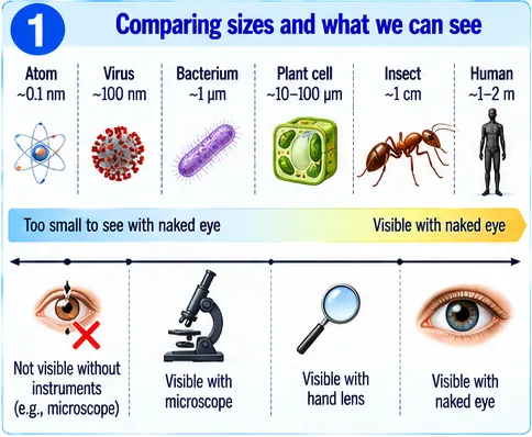
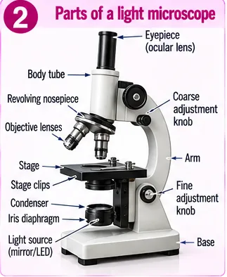
A compound light microscope uses two sets of lenses — the eyepiece (e.g. 10×) and the objective (e.g. 4×, 10×, 40×). Total magnification = eyepiece × objective (10 × 40 = 400×). Thin specimens are stained (iodine for onion peel, methylene blue for cheek cells) to make parts visible. Electron microscopes magnify much more and reveal fine details of organelles.
In 1665 Robert Hooke saw “cells” in cork; Leeuwenhoek (1674) saw living cells in pond water; Robert Brown (1831) discovered the nucleus; Purkinje (1839) coined “protoplasm”.
3. Structure of a Cell
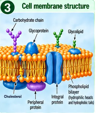
| Feature | Prokaryotic cell | Eukaryotic cell |
|---|---|---|
| Nucleus | No nuclear membrane; DNA in a nucleoid | True nucleus with a nuclear envelope |
| Membrane-bound organelles | Absent | Present (mitochondria, ER, Golgi…) |
| Size | Small (1–10 µm) | Larger (10–100 µm) |
| Examples | Bacteria | Plants, animals, fungi, protists |
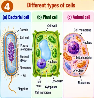
(a) Cell membrane (plasma membrane)
The cell membrane is a thin, flexible boundary made of a phospholipid bilayer with proteins embedded in it — described as a fluid mosaic. Phospholipids have water-loving (hydrophilic) heads facing outwards and water-fearing (hydrophobic) tails facing inwards. Proteins, cholesterol and carbohydrate chains (glycoproteins, glycolipids) help in transport and cell recognition.
The membrane is selectively permeable — it lets some substances pass and stops others. Substances move by:
- Diffusion — from higher to lower concentration (O₂ in, CO₂ out).
- Osmosis — movement of water through a selectively permeable membrane from a dilute to a concentrated solution. A cell in a hypotonic (dilute) solution swells; in a hypertonic (concentrated) solution it shrinks — plant cells show plasmolysis.
- Endocytosis — engulfing food and particles (as in Amoeba).
(b) Cell wall
Plant cells (and fungi and bacteria) have a rigid cell wall outside the membrane. In plants it is made mainly of cellulose; it gives shape and strength and lets plant cells survive in dilute solutions without bursting.
(c) Nucleus
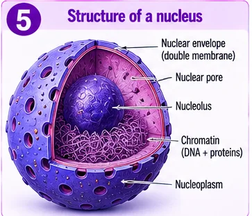
The nucleus is the control centre. It is bounded by a double-layered nuclear envelope with pores, contains the dense nucleolus (makes ribosome parts), and chromatin — DNA with proteins — which condenses into chromosomes during division. DNA carries the information (genes) for making proteins and for inheritance.
4. The Cell Interior — A Coordinated Working System
| Organelle | Structure | Function |
|---|---|---|
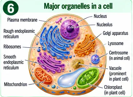
| Endoplasmic reticulum (ER) | Network of membrane tubes; rough ER has ribosomes, smooth ER does not | RER: makes proteins; SER: makes lipids, detoxifies poisons |
| Ribosomes | Tiny granules (no membrane) | Sites of protein synthesis |
| Golgi apparatus | Stacks of flattened sacs | Modifies, packs and dispatches proteins; forms lysosomes |
| Lysosomes | Sacs of digestive enzymes | Digest worn-out parts and germs — “suicide bags” |
| Mitochondria | Double membrane, inner one folded (cristae) | Cellular respiration; release energy as ATP — “powerhouse” |
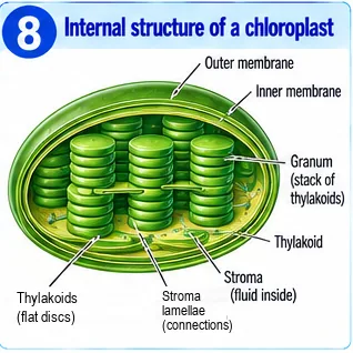
| Plastids | Chloroplasts (green, with thylakoids/grana) and leucoplasts | Photosynthesis; storage of starch, oils |
| Vacuoles | Large central vacuole in plants; small in animals | Store water, salts, wastes; keep plant cells firm (turgid) |
| Centrosome | Two centrioles (animal cells) | Helps in cell division |
Mitochondria and plastids have their own DNA and ribosomes, so they can make some of their own proteins.
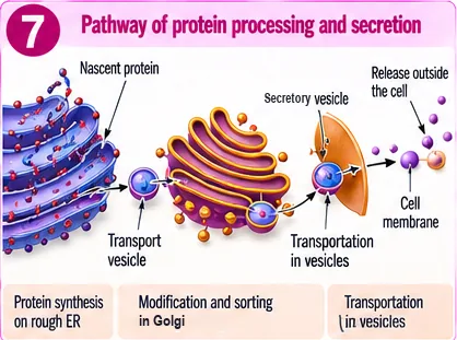
Teamwork: making and sending out a protein
- Instructions in the nucleus (DNA) are copied and sent to ribosomes.
- Ribosomes on the rough ER build the protein.
- Transport vesicles carry it to the Golgi apparatus, where it is modified and sorted.
- Secretory vesicles carry it to the cell membrane and release it outside (e.g. digestive enzymes, hormones).
5. How Do Cells Grow and Divide?
Cells divide to help organisms grow, replace dead cells, repair injuries and reproduce.
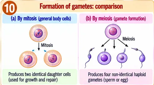
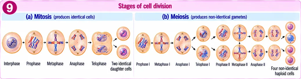
- Mitosis — one cell divides into two identical daughter cells with the same number of chromosomes as the parent. Stages: prophase, metaphase, anaphase, telophase (followed by division of cytoplasm). Used for growth and repair in body cells.
- Meiosis — occurs in reproductive organs; one cell divides twice to form four cells each with half the number of chromosomes; they are not identical. These become gametes (sperm and eggs). When gametes fuse at fertilisation, the full chromosome number is restored.
6. Cell Theory
Proposed by Schleiden (plants, 1838) and Schwann (animals, 1839), and extended by Virchow (1855):
- All living organisms are made of one or more cells.
- The cell is the basic structural and functional unit of life.
- All cells arise from pre-existing cells (omnis cellula e cellula).
Viruses are an exception — they are not made of cells and show life only inside host cells.
7. Master Table: Key Terms and Facts
| Term | Meaning |
|---|---|
| Magnification | Eyepiece power × objective power |
| Prokaryote / Eukaryote | No true nucleus / true nucleus with membrane |
| Selectively permeable | Allows some substances to pass, not others |
| Osmosis | Water moves from dilute to concentrated solution across a membrane |
| Plasmolysis | Shrinking of plant cell contents in a hypertonic solution |
| Chromatin / Chromosome | DNA + protein threads / condensed form during division |
| Mitosis | 2 identical cells, same chromosome number |
| Meiosis | 4 non-identical cells, half chromosome number |
| Cell theory | Schleiden, Schwann, Virchow |
8. Master Summary Table
| Feature | Plant cell | Animal cell |
|---|---|---|
| Cell wall | Present (cellulose) | Absent |
| Plastids | Present | Absent |
| Vacuole | Large central | Small, many or absent |
| Centrosome | Usually absent | Present |
| Shape | Fixed, often rectangular | Irregular or round |
| Lysosomes | Rare | Common |
9. Mind Map
- Light, electron microscope
- Hooke, Leeuwenhoek, Brown
- Fluid mosaic membrane
- Diffusion, osmosis; wall
- Nucleus, ER, Golgi, ribosomes
- Mitochondria, plastids, lysosomes
- Mitosis: 2 identical
- Meiosis: 4 gametes; cell theory
10. Tips and Tricks
- Organelles with their own DNA: “M&P” — Mitochondria and Plastids.
- Protein route: “R-E-G-V-M” — Ribosome, ER, Golgi, Vesicle, Membrane.
- Osmosis: water goes where the solute is more (towards the concentrated side).
- MitOsis = One division, twO identical; MeIosis = makes gametes, halves chromosomes.
- Saying bacteria have no DNA — they have DNA in a nucleoid.
- Saying plant cells have no mitochondria — they have both mitochondria and chloroplasts.
- Confusing cell wall (fully permeable) with cell membrane (selectively permeable).
11. Important One-liners
- Robert Hooke coined the term “cell” (1665).
- Robert Brown discovered the nucleus (1831).
- The cell membrane is selectively permeable.
- The plant cell wall is made of cellulose.
- Mitochondria are the powerhouses of the cell.
- Lysosomes are called suicide bags.
- Ribosomes are the sites of protein synthesis.
- Golgi apparatus packages and dispatches materials.
- Mitosis produces two identical cells; meiosis produces four gametes.
- Virchow stated that all cells arise from pre-existing cells.
12. Exam-pattern Questions with Detailed Answers
Which organelle is known as the powerhouse of the cell?
- Golgi apparatus
- Mitochondrion
- Lysosome
- Ribosome
Show answer
Mitochondria carry out cellular respiration and release energy as ATP. Golgi packs materials, lysosomes digest, ribosomes make proteins.
Answer: (B) Mitochondrion
A microscope has a 10× eyepiece and a 40× objective. What is the total magnification?
Show answer
10 × 40 = 400×.
Answer: 400×
Name two organelles that have their own DNA.
Show answer
Mitochondria and plastids (chloroplasts).
Answer: Mitochondria, plastids
Assertion (A): Plant cells do not burst when placed in pure water. Reason (R): Plant cells have a rigid cell wall.
- Both true; R explains A
- Both true; R does not explain A
- A true, R false
- A false, R true
Show answer
Water enters by osmosis and the cell swells, but the rigid cellulose wall resists further expansion, so the cell becomes turgid without bursting.
Answer: (A)
What happens when a red blood cell is placed in (a) pure water, (b) concentrated salt solution?
Show answer
(a) Water enters by osmosis; the cell swells and may burst, as animal cells have no wall. (b) Water leaves the cell; it shrinks.
Answer: Swells/bursts; shrinks
Give three differences between prokaryotic and eukaryotic cells.
Show answer
Prokaryotes: no nuclear membrane (nucleoid), no membrane-bound organelles, smaller size (bacteria). Eukaryotes: true nucleus, membrane-bound organelles, larger (plants, animals).
Answer: Nucleus, organelles, size
Why are lysosomes called suicide bags?
Show answer
They contain powerful digestive enzymes. When a cell is damaged, lysosomes may burst and their enzymes digest the cell itself.
Answer: Their enzymes can digest their own cell
Trace the path of a digestive enzyme from its synthesis to its release from a cell.
Show answer
Made by ribosomes on the rough ER → carried in transport vesicles to the Golgi apparatus → modified and packed → secretory vesicles carry it to the cell membrane → released outside by fusion with the membrane.
Answer: RER → vesicle → Golgi → secretory vesicle → membrane → outside
Why is meiosis necessary for sexual reproduction?
Show answer
Gametes must have half the chromosomes so that when two fuse at fertilisation, the zygote has the normal number (e.g. 23 + 23 = 46 in humans). Without meiosis, chromosome number would double every generation. Meiosis also creates variation.
Answer: Keeps chromosome number constant across generations; adds variation
Case-based: Raisins kept in water swell; fresh grapes kept in strong sugar syrup shrink. (a) Name the process. (b) Explain both. (c) Which property of the membrane makes this possible?
Show answer
(a) Osmosis. (b) Raisins have a concentrated interior, so water enters and they swell; syrup is more concentrated than grapes, so water leaves and they shrink. (c) The membrane is selectively permeable — water passes but sugar does not easily.
Answer: Osmosis; water moves towards higher concentration; selective permeability
Explain the fluid mosaic nature of the cell membrane.
Show answer
The membrane is a phospholipid bilayer in which lipid molecules can move sideways (fluid), and proteins of different kinds are embedded or attached like tiles in a mosaic. This flexible structure allows the membrane to change shape, fuse with vesicles and regulate transport.
Answer: Moving lipid bilayer with scattered proteins
A cell lacks a nucleus, cell wall and mitochondria but is found in human blood and carries oxygen. Identify it and explain how it survives without these.
Show answer
Mature human red blood cell. Losing the nucleus and organelles gives more space for haemoglobin to carry oxygen. It gets energy by breaking glucose without oxygen and lives only about 120 days, after which new RBCs from bone marrow replace it.
Answer: Red blood cell — specialised for oxygen transport, short-lived
13. Quick Revision Summary
- Cells are tiny; microscopes (magnification = eyepiece × objective) reveal them.
- Prokaryotes lack a true nucleus; eukaryotes have one and membrane-bound organelles.
- Membrane: fluid mosaic, selectively permeable — diffusion, osmosis, endocytosis; plants have cellulose walls.
- Nucleus controls; ER, ribosomes, Golgi, lysosomes, mitochondria, plastids, vacuoles each have roles and work together.
- Mitosis → 2 identical cells; meiosis → 4 gametes with half chromosomes. Cell theory: Schleiden, Schwann, Virchow.